File:DaviesHarding1944 fig01.jpg

Original file (979 × 1,000 pixels, file size: 241 KB, MIME type: image/jpeg)
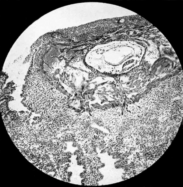
Fig 1. Ovum and Endometrium
Operculum, consisting of fibrin and leucocytes permeated by plasmoditrophoblast, is seen in upper part of photograph. Fibrinous wisp projects into mouth of gland on left. No epithelium on surface of operculum. Sprouts of trophoblast seen penetrating stratum compactum below ovum. Maternal red-blood corpuscles seen in lucunae of plasmoditrophoblast to left of chorionic vesicle. Primitive mesoblast shown permeating chorionic vesicle between cytotrophoblast. and thin exocoelomic membrane. Of the two cavities inside the chorionic.vesicle, the upper, larger one is the exocoelomic vesicle (primitive yolk sac), with the endodermal plate of cubical cells forming its lower wall.
The lower, smaller cavity in the amniotic cavity; in the picture its upper wall is formed by the columnar cells of the ectodermal disc. The endometrium adjacent to the trophoblast shows no zone of necrosis. x 90.
| Historic Disclaimer - information about historic embryology pages |
|---|
| Pages where the terms "Historic" (textbooks, papers, people, recommendations) appear on this site, and sections within pages where this disclaimer appears, indicate that the content and scientific understanding are specific to the time of publication. This means that while some scientific descriptions are still accurate, the terminology and interpretation of the developmental mechanisms reflect the understanding at the time of original publication and those of the preceding periods, these terms, interpretations and recommendations may not reflect our current scientific understanding. (More? Embryology History | Historic Embryology Papers) |
| Stage 5 Links: Week 2 | Implantation | Lecture | Practical | Carnegie Embryos | Category:Carnegie Stage 5 | Next Stage 6 | ||
| Historic Papers: 1941 | 1944 day 9-10 | 1945 day 7.5 | 1945 day 9-10 | ||
|
Reference
Davies F. and Harding HE. A Human ovum nine to ten days old. (1944) BJOG: An International Journal of Obstetrics & Gynaecology 51(3): 225-230.
Cite this page: Hill, M.A. (2024, April 27) Embryology DaviesHarding1944 fig01.jpg. Retrieved from https://embryology.med.unsw.edu.au/embryology/index.php/File:DaviesHarding1944_fig01.jpg
- © Dr Mark Hill 2024, UNSW Embryology ISBN: 978 0 7334 2609 4 - UNSW CRICOS Provider Code No. 00098G
File history
Click on a date/time to view the file as it appeared at that time.
| Date/Time | Thumbnail | Dimensions | User | Comment | |
|---|---|---|---|---|---|
| current | 19:05, 5 August 2016 |  | 979 × 1,000 (241 KB) | Z8600021 (talk | contribs) | |
| 19:03, 5 August 2016 |  | 1,200 × 1,636 (438 KB) | Z8600021 (talk | contribs) | Fig 1. Ovum and endometrium. Operculum, consisting of fibrin and leucocytes permeated by plasmoditrophoblast, is seen in upper part of photograph. Fibrinous wisp projects into mouth of gland on left. No epithelium on surface of operculum. Sprouts of t... |
You cannot overwrite this file.
File usage
The following page uses this file:
