File:Corpus luteum.jpg
Corpus_luteum.jpg (450 × 600 pixels, file size: 94 KB, MIME type: image/jpeg)
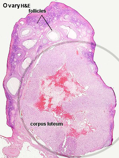
Ovary - Corpus Luteum
Histology image shows the corpus luteum (CL, yellow body)
- The ovulating follicle forms initially the corpus hemorrhagicum and then corpus luteum.
- Corpus luteum (CL, yellow body) outer layer consisting of theca lutein cells and more centrally the granulosa lutein cells.
- Corpus luteum core is initially filled with a blood clot, following ovulation, and is the invaded by connective tissue.
Theca lutein cells and granulosa lutein cells work together in the production of ovarian hormones that support the initial pregnancy. Lack of implantation and associated hCG will lead to this structure not producing hormones and forming the corpus albicans (white body).
Theca Lutein Cells
- darker stained cells.
- derived from the theca interna of the original follicle.
- lack microvilli on the surface.
- lack the aromatase enzyme.
- produce androgens for the granulosa lutein cells to convert.
Granulosa Lutein Cells
- lighter stained cells.
- derived from the granulosa cells of the original follicle.
- contain aromatase enzyme.
- produce estrogen and progesterone from the androgens produced by the theca lutein cells.
Links: Histology | Histology Stains | Blue Histology images copyright Lutz Slomianka 1998-2009. The literary and artistic works on the original Blue Histology website may be reproduced, adapted, published and distributed for non-commercial purposes. See also the page Histology Stains.
Cite this page: Hill, M.A. (2024, April 27) Embryology Corpus luteum.jpg. Retrieved from https://embryology.med.unsw.edu.au/embryology/index.php/File:Corpus_luteum.jpg
- © Dr Mark Hill 2024, UNSW Embryology ISBN: 978 0 7334 2609 4 - UNSW CRICOS Provider Code No. 00098G
Histology image H&E high power Clu01he.jpg
File history
Click on a date/time to view the file as it appeared at that time.
| Date/Time | Thumbnail | Dimensions | User | Comment | |
|---|---|---|---|---|---|
| current | 16:58, 6 May 2012 | | 450 × 600 (94 KB) | Z8600021 (talk | contribs) | |
| 10:14, 3 August 2009 | 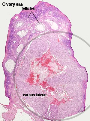 | 300 × 400 (55 KB) | MarkHill (talk | contribs) | Corpus luteum Histology image H&E low power Image Source: Lutz Slomianka, UWA Blue Histology Clu01he.jpg http://www.lab.anhb.uwa.edu.au/mb140/CorePages/FemaleRepro/femalerepro.htm#Corpus |
You cannot overwrite this file.
File usage
The following 11 pages use this file:
- 2010 BGD Practical 3 - Implantation
- 2011 Lab 2 - Week 2
- ANAT2241 Female Reproductive System
- ANAT2341 Lab 2 - Week 2
- BGDA Practical - Female Reproductive Tract Histology
- BGDA Practical 3 - Implantation
- Corpus Luteum Development
- Endocrine System Development
- Human Chorionic Gonadotropin
- Ovary Development
- Talk:2011 Lab 2 - Week 2