File:Chicken-neural-crest-migration-04.jpg
Chicken-neural-crest-migration-04.jpg (600 × 397 pixels, file size: 67 KB, MIME type: image/jpeg)
Neural crest migration
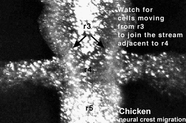
Chicken embryo sequence sequence shows the migration of DiI-labeled neural crest cells from r3 follow a caudolateral trajectory to join cells exiting adjacent to r4.
r = rhombomere
Duration: 4 hrs Time interval between images: 3 min
Each image represents 10 confocal sections separated by 10 microns each, projected onto 1 image.
Links: Movies - Chicken Neural Crest | Neural Crest Development | File:Chicken-neural_crest_migration_04.flv | all Development movies
<pubmed>10683170</pubmed>
Original Neural Crest movies kindly provided by Paul Kulesa
Reproduced with Permission Development, © The Company of Biologists Ltd. 2000.
File history
Click on a date/time to view the file as it appeared at that time.
| Date/Time | Thumbnail | Dimensions | User | Comment | |
|---|---|---|---|---|---|
| current | 12:31, 12 August 2010 |  | 600 × 397 (67 KB) | S8600021 (talk | contribs) | ==Neural crest migration== Chicken embryo sequence sequence shows the migration of DiI-labeled neural crest cells from r3 follow a caudolateral trajectory to join cells exiting adjacent to r4. r = rhombomere '''Links:''' [[Movies - Chi |
You cannot overwrite this file.
File usage
The following 26 pages use this file:
- 2010 Lab 3
- 2010 Lecture 12
- 2011 Lab 3 - Week 4
- ANAT2341 Lab 3 - Week 4
- Chicken Development
- Chicken Neural Crest Migration Movie 1
- Chicken Neural Crest Migration Movie 2
- Chicken Neural Crest Migration Movie 3
- Chicken Neural Crest Migration Movie 4
- Chicken Neural Crest Migration Movie 5
- Chicken Neural Crest Migration Movie 6
- Chicken Neural Crest Migration Movie 7
- Developmental Mechanisms
- Lecture - Neural Crest Development
- Movie - Chicken Neural Crest Migration 01
- Movies
- Movies - Chicken Neural Crest
- Neural Crest - Cranial Nerve Development
- Neural Crest - Cranial Nerves
- Neural Crest Development
- Talk:2011 Lab 3
- Talk:Flash Movies
- Talk:Quicktime Movies
- Template:Chicken neural crest movies
- Template:Neural Crest movie 4
- Template talk:Chicken neural crest movies
