File:Brain fissure development 01.jpg

Original file (1,157 × 502 pixels, file size: 47 KB, MIME type: image/jpeg)
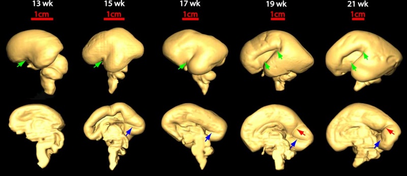
Brain Development
Three-dimensional reconstruction of the lateral (top row) and medial (bottom row) surface of 13–21 week brains to reveal the development of the Sylvian fissure (green arrow), the calcarine fissure (blue arrow), and the parieto-occipital sulcus (red arrow), respectively.
Original File Name: Figure 5
Reference
<pubmed>19339620</pubmed>| PMC2721010 | J Neurosci.
Copyright: Copyright of all material published in The Journal of Neuroscience remains with the authors. The authors grant the Society for Neuroscience an exclusive license to publish their work for the first 6 months. After 6 months the work becomes available to the public to copy, distribute, or display under a Creative Commons Attribution-Noncommercial-Share Alike 3.0 Unported license.
File history
Click on a date/time to view the file as it appeared at that time.
| Date/Time | Thumbnail | Dimensions | User | Comment | |
|---|---|---|---|---|---|
| current | 11:20, 27 August 2010 | | 1,157 × 502 (47 KB) | S8600021 (talk | contribs) | ==Brain Development== Three-dimensional reconstruction of the lateral (top row) and medial (bottom row) surface of 13–21 week brains to reveal the development of the Sylvian fissure (green arrow), the calcarine fissure (blue arrow), and the parieto-occ |
You cannot overwrite this file.
File usage
The following 2 pages use this file: