File:Bone marrow histology 01.jpg: Difference between revisions
From Embryology
(==Bone Marrow Histology== Bone marrow histology 01.jpg {{Bone Marrow Histology}} {{Blue Histology}}) |
|||
| (10 intermediate revisions by the same user not shown) | |||
| Line 1: | Line 1: | ||
==Bone Marrow Histology== | ==Bone Marrow Histology== | ||
Bone marrow histology | * Overview of bone marrow cells and structure. | ||
* Marrow contains a large number of developing and mature blood cells at various stages of differentiation and appearance. | |||
** Histology students always make the mistake of trying to identify all the cell types. | |||
* '''parenchyma''' - haematopoietic (hematopoietic) cell compartment. | |||
* '''stroma''' - fibroblasts, adipocytes, nerves, and the bone marrow’s vascular system. | |||
See also the matching field image showing [[:File:Bone marrow histology 02.jpg|megakaryoblast and megakaryocyte]]. | |||
===Sinusoid=== | |||
* are supplied by arterioles and capillaries vessels spanning throughout the bone marrow. | |||
* wall consists of a single layer of endothelial cells and no supporting cells. | |||
* interconnected by intersinusoidal capillaries. | |||
* radially distributed around the draining central sinus (about 100 µm in diameter). | |||
* bone marrow sinusoids are unique and are not comparable with regular veins. | |||
* see [[File_talk:Bone_marrow_histology_01.jpg|Bone Marrow Vascular Niche]] | |||
===Adipocyte=== | |||
* fat cells. | |||
* large cell size with peripheral flattened nucleus. | |||
* enclose open spaces that were occupied by lipid droplets. | |||
** lipid is lost during histological processing. | |||
* number correlates inversely with bone marrow haematopoietic activity. | |||
** more adipocytes (yellow marrow) less haematopoiesis. | |||
** less adipocytes (red marrow) more haematopoiesis. | |||
* recently identified as a negative regulator of the haematopoietic microenvironment PMID 19516257 | |||
* See also [[Endocrine_-_Other_Tissues#Adipose_Tissue|Endocrine - Adipose Tissue]] | |||
{{Bone Marrow Histology}} | {{Bone Marrow Histology}} | ||
{{Blue Histology}} | {{Blue Histology}} | ||
Latest revision as of 16:21, 18 February 2019
Bone Marrow Histology
- Overview of bone marrow cells and structure.
- Marrow contains a large number of developing and mature blood cells at various stages of differentiation and appearance.
- Histology students always make the mistake of trying to identify all the cell types.
- parenchyma - haematopoietic (hematopoietic) cell compartment.
- stroma - fibroblasts, adipocytes, nerves, and the bone marrow’s vascular system.
See also the matching field image showing megakaryoblast and megakaryocyte.
Sinusoid
- are supplied by arterioles and capillaries vessels spanning throughout the bone marrow.
- wall consists of a single layer of endothelial cells and no supporting cells.
- interconnected by intersinusoidal capillaries.
- radially distributed around the draining central sinus (about 100 µm in diameter).
- bone marrow sinusoids are unique and are not comparable with regular veins.
- see Bone Marrow Vascular Niche
Adipocyte
- fat cells.
- large cell size with peripheral flattened nucleus.
- enclose open spaces that were occupied by lipid droplets.
- lipid is lost during histological processing.
- number correlates inversely with bone marrow haematopoietic activity.
- more adipocytes (yellow marrow) less haematopoiesis.
- less adipocytes (red marrow) more haematopoiesis.
- recently identified as a negative regulator of the haematopoietic microenvironment PMID 19516257
- See also Endocrine - Adipose Tissue
- Bone Marrow Histology: Blood Development | Marrow overview | Megakaryocyte | Megakaryocyte detail | Myelocyte | Normoblast | Reticulocyte | Blood Histology | Bone Development | Category:Blood
Links: Histology | Histology Stains | Blue Histology images copyright Lutz Slomianka 1998-2009. The literary and artistic works on the original Blue Histology website may be reproduced, adapted, published and distributed for non-commercial purposes. See also the page Histology Stains.
Cite this page: Hill, M.A. (2024, May 19) Embryology Bone marrow histology 01.jpg. Retrieved from https://embryology.med.unsw.edu.au/embryology/index.php/File:Bone_marrow_histology_01.jpg
- © Dr Mark Hill 2024, UNSW Embryology ISBN: 978 0 7334 2609 4 - UNSW CRICOS Provider Code No. 00098G
File history
Click on a date/time to view the file as it appeared at that time.
| Date/Time | Thumbnail | Dimensions | User | Comment | |
|---|---|---|---|---|---|
| current | 07:27, 25 February 2012 | 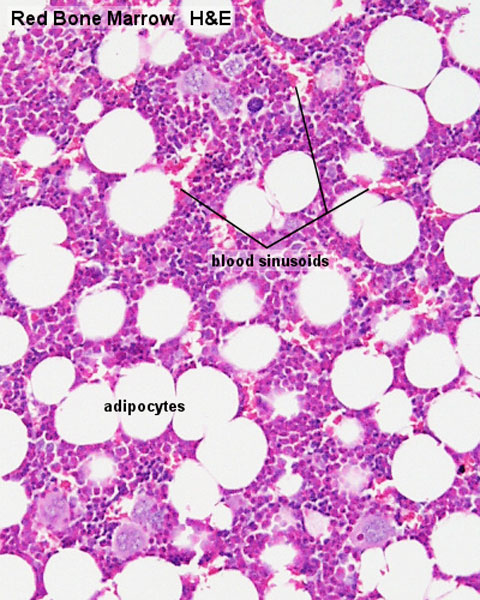 | 480 × 600 (114 KB) | Z8600021 (talk | contribs) | ==Bone Marrow Histology== Bone marrow histology 01.jpg {{Bone Marrow Histology}} {{Blue Histology}} |
You cannot overwrite this file.