File:Bartelmez1923 fig02.jpg

Original file (1,295 × 2,189 pixels, file size: 218 KB, MIME type: image/jpeg)
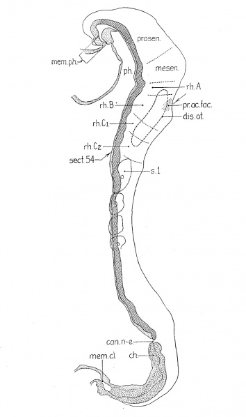
Fig. 2. A reconstruction of a four—somite embryo
(H279, U. of C. Coll.; Carnegie 3709); magnified 200 diameters and reduced one half in reproduction. The plane of section is indicated by the position of section 54 which was shown as figure 1 in my 1922 paper.
camn-e., neurenteric canal; ch., chorda; disc.o£., otic disc; mom. cl., cloacal membrane (the entoderm has shrunken away from the cctoderm) ; mem. ph., pharyngeal membrane; mesen., midbrain; ph., pharynx; pr.ac.fac., acoustic0-facial primordium; prosen., forebrain; rh.A, rh.B, first and second hindbrain segments; rh..C'1 and 7'h.C'2 have resulted from the division of rh.C’ in the previous embryo; rh.B is identical with the definitive fourth rhombomere, rh.C'; with the fifth.
Figure 2 gives the relations in a four-somite embryo (H279, U. of C. Coll.; Carnegie 3709) in which abundant mitoses are to be seen, although the specimen underwent great shrinkage during dehydration. It belongs to a group which includes that just described as well as Wilson’s ‘H3’ (’14) and ‘Klb’ (Keibel and Elze, ’O8, Normentafel No. 3). Forebrain, midbrain, and three hindbrain subdivisions were clearly visible in the gross specimen when studied in 80 per cent alcohol. It will be seen in the figure that the forebrain has begun to grow forward beyond the pharynx and that it is relatively longer than in the previous case. The midbrain is wedge—shaped. There are clear signs of differentiation in the hindbrain in that there are four segments and a ganglionic anlage. The first segment (rh.A) is still small and inconspicuous, whereas the second (rh.B) has acquired new characters which distinguish it for the rest of our period. The enlargement is sharply marked off by constrictions fore and aft, and corresponding to it there is a dip in the floor. Ventrally the neural groove is enlarged in the manner typical for neuromeres, but dorsally the folds approach one another only to diverge again at the dorsal edge where they are thickened. The thickening is the acoustico-facial primordium (pr.ac.fac.). These relations may be seen in figure 1 of my 1922 paper. The level of that section is indicated in the present figure 2, where it will be seen that the plane of section passes obliquely through the otic segment (rh.B) and the two succeeding segments. The latter two (rh.C1 and rh.C2) correspond in position to the third hindbrain segment of the previous specimen (rh.C). It may be that that segment has divided, but it is possible that H279 is an individual in which the segmentation of the nervous system is particularly pronounced.
Online Editor Embryo H279 (University of Chicago Collection) became Carnegie Collection Embryo no. 3709, 4 somite embryo corresponds to Carnegie stage 10 in Week 4.
| Historic Disclaimer - information about historic embryology pages |
|---|
| Pages where the terms "Historic" (textbooks, papers, people, recommendations) appear on this site, and sections within pages where this disclaimer appears, indicate that the content and scientific understanding are specific to the time of publication. This means that while some scientific descriptions are still accurate, the terminology and interpretation of the developmental mechanisms reflect the understanding at the time of original publication and those of the preceding periods, these terms, interpretations and recommendations may not reflect our current scientific understanding. (More? Embryology History | Historic Embryology Papers) |
Reference
Bartelmez GW. The subdivisions of the neural folds in man. (1923) J. Comp. Neural., 35: 231-247.
Cite this page: Hill, M.A. (2024, April 27) Embryology Bartelmez1923 fig02.jpg. Retrieved from https://embryology.med.unsw.edu.au/embryology/index.php/File:Bartelmez1923_fig02.jpg
- © Dr Mark Hill 2024, UNSW Embryology ISBN: 978 0 7334 2609 4 - UNSW CRICOS Provider Code No. 00098G
File history
Click on a date/time to view the file as it appeared at that time.
| Date/Time | Thumbnail | Dimensions | User | Comment | |
|---|---|---|---|---|---|
| current | 22:40, 7 June 2016 |  | 1,295 × 2,189 (218 KB) | Z8600021 (talk | contribs) | ==Fig. 2 A reconstruction of a four—somite embryo== (H279, U. of C. Coll.); magnified 200 diameters and reduced one half in reproduction. The plane of section is indicated by the position of section 54 which was shown as figure 1 in my 1922 paper. c... |
You cannot overwrite this file.
File usage
The following 2 pages use this file:
