File:Bartelmez1923 fig01.jpg

Original file (1,312 × 1,919 pixels, file size: 245 KB, MIME type: image/jpeg)
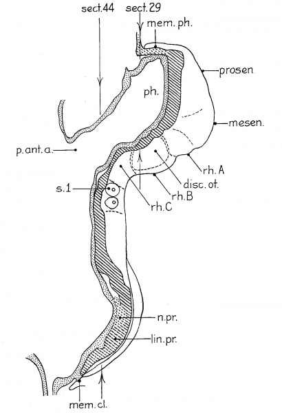
Fig. 1. A projection reconstruction on the midsagittal plane of a two-somite embryo
(Embryo no. 1878, Carnegie Collection). x100. The cut surfaces of the nervous system are hatched, those of the gut, stippled. The body ectoderm and the primitive streak are indicated by broken lines. The arrows show the plane of section. Section 44 is reproduced by Ingalls (’20) as figure A, page 81. disc.ot., otic disc or plate; lm.pr., primitive streak; mem.cl., cloacal membrane; mem.ph., pharyngeal membrane; mesen.,midbrain; n.pr., primitive node of Hansen; p.int.a., anterior intestinal portal; ph., pharynx; prosen., forebrain; rh.A,B, and C’, first, second and third hindbrain segments; 3.1, first somite with myocoale.
Figure 1 presents an analysis of a two—somite embryo (1878 of the Carnegie Collection) which has been well described by Ingalls (’20). The neural folds can be divided into five segments which are separated either by constrictions or by sulci on the lateral surface of the folds. The midbrain (mesen.), defined by the cranial flexure, separates the forebrain (prosen.) from the hindbrain. The latter exhibits three subdivisions: the first (rh.A) is small but clearly defined, especially on the right side; the second, the otic (rh.B), is prominent, and the third (rh.C) involves the rest of the neural folds rostral to the first pair of somites. The otic segment in this specimen differs from those of all succeeding stages in that there is a general enlargement of the neural groove corresponding to it. The otic plate (disc.ot.) is practically coextensive with the otic segment and is well marked, as was pointed out by Ingalls (’20).
Online Editor - Carnegie Collection Embryo no. 1878 corresponds to Carnegie stage 9 occurring during Week 3.
Ingalls NW. A human embryo at the beginning of segmentation, with special reference to the vascular system. (1920) Contrib. Embryol., Carnegie Inst. Wash. Publ. 274, 11: 61-90.
| Historic Disclaimer - information about historic embryology pages |
|---|
| Pages where the terms "Historic" (textbooks, papers, people, recommendations) appear on this site, and sections within pages where this disclaimer appears, indicate that the content and scientific understanding are specific to the time of publication. This means that while some scientific descriptions are still accurate, the terminology and interpretation of the developmental mechanisms reflect the understanding at the time of original publication and those of the preceding periods, these terms, interpretations and recommendations may not reflect our current scientific understanding. (More? Embryology History | Historic Embryology Papers) |
Reference
Bartelmez GW. The subdivisions of the neural folds in man. (1923) J. Comp. Neural., 35: 231-247.
Cite this page: Hill, M.A. (2024, April 27) Embryology Bartelmez1923 fig01.jpg. Retrieved from https://embryology.med.unsw.edu.au/embryology/index.php/File:Bartelmez1923_fig01.jpg
- © Dr Mark Hill 2024, UNSW Embryology ISBN: 978 0 7334 2609 4 - UNSW CRICOS Provider Code No. 00098G
File history
Click on a date/time to view the file as it appeared at that time.
| Date/Time | Thumbnail | Dimensions | User | Comment | |
|---|---|---|---|---|---|
| current | 22:31, 7 June 2016 |  | 1,312 × 1,919 (245 KB) | Z8600021 (talk | contribs) | {{Historic Disclaimer}} ===Reference=== {{Ref-Bartelmez1923}} {{Footer}} |
You cannot overwrite this file.
File usage
The following 4 pages use this file:
