File:Bartelmez1922-fig02.jpg
Original file (1,203 × 1,700 pixels, file size: 317 KB, MIME type: image/jpeg)
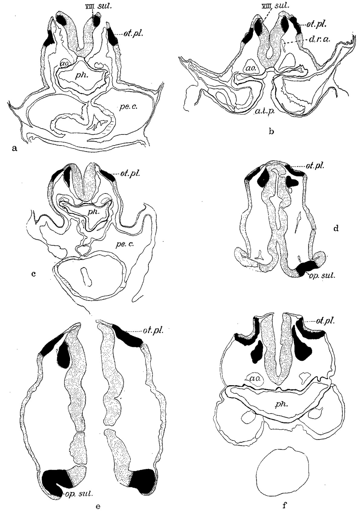
Fig. 2. A series of tracings made from the sections with the Edinger apparatus
X 200, and reduced to 66% diameters in reproduction. The solid color marks the otic plate and the acousticofaeial ganglion and optic anlage; the rest of the nervous system, including the neural crest and the ectoderm, are stippled, the outer boundary of the pharynx is indicated by a heavy line and the myoepicardium marked by horizontal lines. The mesenchyme is not shown except Where it was shrunken and then its outer limits are drawn.
a. Mall embryo no. 391 — eight somites—section 45.
b. H87 — eight somites section 64 (cf. figs. 3 and 4).
c. Eternod ‘Du Ga’ — nine somites — section 49.
d. N.Y.U. embryo no. 4 — fourteen somites — section 32.
e. Pfannenstiel III — fourteen somites — section 41.
f. Mall embryo no. 470 - sixteen somites — s1. 2-1-7.
a.i.p., anterior intestinal portal; ao., aorta; d.r.v., dorsal second aortic ramus; op.sul., optic sulcus; pe.c., pericardial cavity; ph., pharynx; VIII sul.; otic sulcus. In this and all other figures of sections the right side of the embryo appears at the observer’s left.
| Historic Disclaimer - information about historic embryology pages |
|---|
 |
- Links: Fig 1 | Fig 2 | Fig 3 | Fig 4 | Fig 5 | Fig 6 | Fig 7 | Fig 8 | Fig 9 | Fig 10 | Bartelmez 1922 | Historic Embryology Papers | Hearing | Vision
Reference
Bartelmez GW. The origin of the otic and optic primordia in man. (1922) J. Comp. Neural., 34: 201-232.
Cite this page: Hill, M.A. (2024, April 27) Embryology Bartelmez1922-fig02.jpg. Retrieved from https://embryology.med.unsw.edu.au/embryology/index.php/File:Bartelmez1922-fig02.jpg
- © Dr Mark Hill 2024, UNSW Embryology ISBN: 978 0 7334 2609 4 - UNSW CRICOS Provider Code No. 00098G
File history
Click on a date/time to view the file as it appeared at that time.
| Date/Time | Thumbnail | Dimensions | User | Comment | |
|---|---|---|---|---|---|
| current | 00:09, 19 September 2015 | | 1,203 × 1,700 (317 KB) | Z8600021 (talk | contribs) | |
| 00:08, 19 September 2015 |  | 1,352 × 2,392 (607 KB) | Z8600021 (talk | contribs) |
You cannot overwrite this file.
File usage
The following page uses this file: