File:Bailey468.jpg
Bailey468.jpg (774 × 519 pixels, file size: 60 KB, MIME type: image/jpeg)
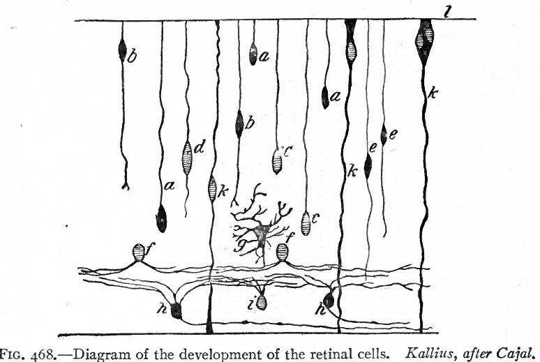
Fig. 468. Diagram of the development of the retinal cells
Kallius, after Cajal.
- a, Cone cells in unipolar stage
- fe, cone cells in bipolar stage
- c, rod cells in unipolar stage
- d, rod cells in bipolar stage
- e, bipolar cells
- i, amacrine cells
- g, horizontal cell
- h, ganglion cells
- k, Muller's cells or fibbers
- l, external limiting membrane.
About the end of the eighth week the inner part of the primitive nuclear layer differentiates into the layer of eanzlion cetts (Fig. 468, h). These are large cells and with their processes constitute the third or proximal optic neurone. They can be first distinguished in the fundus of the cup and gradually extend to the ora serrata. They are the first of the cellular dements of the adult retina which can be definitely recognized as such. From each cell, two kinds of processes develop, dendrites, which ramify in this and in the more external layers of the retina, and an axone which grows toward the cavity of the eye and becomes a fiber of the layer of nerve fibers, whence it continues into the optic stalk as one of the fibers of the optic nerve. The layer of ganglion cells is thickest in an area situated somewhat lateral to the attachment of the optic stalk and known as the area centralis. It is distinguishable about the end of the fourth month. In the center of the area centralis the retinal layers become thin to form the fovea centralis which develops toward the end of foetal life. The macula lutea with its yellow pigment does not develop until after birth. The retina at this stage thus consists of four layers which from within outward are (i) the layer of nerve fibers, (2) the layer of ganglion cells, (3) the nuclear layer, (4) the pigmented layer (see Fig. 469).
- Text-Book of Embryology: Germ cells | Maturation | Fertilization | Amphioxus | Frog | Chick | Mammalian | External body form | Connective tissues and skeletal | Vascular | Muscular | Alimentary tube and organs | Respiratory | Coelom, Diaphragm and Mesenteries | Urogenital | Integumentary | Nervous System | Special Sense | Foetal Membranes | Teratogenesis | Gallery of All Figures
| Historic Disclaimer - information about historic embryology pages |
|---|
 |
Reference
Bailey FR. and Miller AM. Text-Book of Embryology (1921) New York: William Wood and Co.
Cite this page: Hill, M.A. (2024, April 27) Embryology Bailey468.jpg. Retrieved from https://embryology.med.unsw.edu.au/embryology/index.php/File:Bailey468.jpg
- © Dr Mark Hill 2024, UNSW Embryology ISBN: 978 0 7334 2609 4 - UNSW CRICOS Provider Code No. 00098G
File history
Click on a date/time to view the file as it appeared at that time.
| Date/Time | Thumbnail | Dimensions | User | Comment | |
|---|---|---|---|---|---|
| current | 13:48, 1 February 2011 | | 774 × 519 (60 KB) | S8600021 (talk | contribs) |
You cannot overwrite this file.