File:Bailey467.jpg
Original file (946 × 515 pixels, file size: 159 KB, MIME type: image/jpeg)
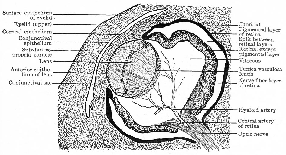
Fig. 467. Horizontal section through eye of human embryo of 13-14 weeks
Modified from Lange.
In man and in Mammals generally it is more or less filled with cells. These, however, degenerate and take no part in the formation of the permanent lens. Comparing the posterior with the anterior wall of the lens at this stage, the latter is seen to be composed of a single layer of cuboidal cells, the anlage of the anterior epithelium of the lens (Figs. 463, 465, g, h, i). This layer passes over rather abruptly into the posterior wall which consists of a single layer of greatly elongated lens cells, the anlagen of the lens fibers. The lens fibers continue to elongate until by the end of the second month they touch the anterior epithelium, thus completely obliterating the cavity of the lens vesicle (Fig. 467). A small cleft containing a few drops of fluid, the liquor Morgagni, may remain between the anterior epithelium and the lens fibers.
By the extension of mesodermic tissue in between the lens and the surface ectoderm, the lens becomes by the end of the sixth week completely surrounded by a layer of vascular connective tissue. This is known as the tunica lentis, and receives its blood supply mainly from the hyaloid artery (Fig. 467) which is a foetal continuation of the arteria centralis retina (p. 545). Branches from the hyaloid artery break up into a capillary network which covers both anterior and posterior surfaces of the lens. That part of the tunica vasculosa which covers the anterior surface of the lens is known as the membrana pupillaris. After the earlier and more rapid formation of lens fibers ceases, the hyaloid artery begins (about the seventh month) to undergo regressive changes, and at birth is normally absent. Rarely more or less of the tunica vasculosa fails to degenerate, and if the part which persists is the membrana pupillaris there results a malformation known as congenital atresia of the pupil.
- Text-Book of Embryology: Germ cells | Maturation | Fertilization | Amphioxus | Frog | Chick | Mammalian | External body form | Connective tissues and skeletal | Vascular | Muscular | Alimentary tube and organs | Respiratory | Coelom, Diaphragm and Mesenteries | Urogenital | Integumentary | Nervous System | Special Sense | Foetal Membranes | Teratogenesis | Gallery of All Figures
| Historic Disclaimer - information about historic embryology pages |
|---|
 |
Reference
Bailey FR. and Miller AM. Text-Book of Embryology (1921) New York: William Wood and Co.
Cite this page: Hill, M.A. (2024, April 26) Embryology Bailey467.jpg. Retrieved from https://embryology.med.unsw.edu.au/embryology/index.php/File:Bailey467.jpg
- © Dr Mark Hill 2024, UNSW Embryology ISBN: 978 0 7334 2609 4 - UNSW CRICOS Provider Code No. 00098G
File history
Click on a date/time to view the file as it appeared at that time.
| Date/Time | Thumbnail | Dimensions | User | Comment | |
|---|---|---|---|---|---|
| current | 13:47, 1 February 2011 | | 946 × 515 (159 KB) | S8600021 (talk | contribs) |
You cannot overwrite this file.