File:Bailey465.jpg

Original file (806 × 931 pixels, file size: 142 KB, MIME type: image/jpeg)
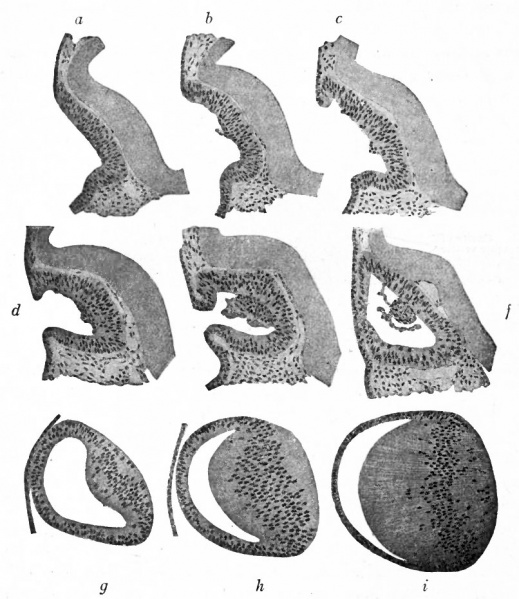
Fig. 465. Successive stages in the development of the lens in the rabbit embryo
Rabl.
- a-e - are from embryos of from 11.5 to 12 days
- f - at end of 12th day
- g - during the 13th day
- h - between the 13th and 14th days
- i - from an embryo of 11 mm.
The lens area is thicker at its center than at its periphery and when the center of the lens area becomes the bottom of the lens depression and later the posterior wall of the lens vesicle this greater thickness is maintained. In fact, the posterior wall of the vesicle becomes still thicker so that it projects into the cavity of the lens vesicle as an eminence (Fig. 465, g.). In the chick the lens vesicle is hollow. In man and in Mammals generally it is more or less filled with cells. These, however, degenerate and take no part in the formation of the permanent lens. Comparing the posterior with the anterior wall of the lens at this stage, the latter is seen to be composed of a single layer of cuboidal cells, the anlage of the anterior epithelium of the lens (Figs. 463, 465, g, h, i).
When the lens fibers are first formed, the longest fibers are in the center and the fibers gradually get shorter toward the periphery of the lens where they pass over into the anterior epithelium (Fig. 465), As the lens develops, the peripheral fibers elongate more rapidly than the central, with the result that in the fully developed lens the central fibers are the shortest, forming a sort of core around which the now longer peripheral fibers extend in much the same manner as the layers of an onion (Fig. 467). The ends of the fibers meet on the anterior and posterior surfaces of the lens, along more or less definite lines which can be seen on surface examination and which are known as sutural lines. The lens fibers are at first all nucleated and as the nuclei are situated at approximately the same level in all the fibers, there results a so-called nuclear zone (Fig. 465, i). Later the nuclei disappear. The sutural lines become evident about the fifth month and mark the completion of the lens formation, although lens fibers continue to be formed throughout fcetal and in postnatal life, probably by proliferation and differentiation of the cells of the anterior epithelium, in the region where the latter pass over into the lens fibers. (The successive stages in the development of the lens are shown in Fig. 465.)
- Text-Book of Embryology: Germ cells | Maturation | Fertilization | Amphioxus | Frog | Chick | Mammalian | External body form | Connective tissues and skeletal | Vascular | Muscular | Alimentary tube and organs | Respiratory | Coelom, Diaphragm and Mesenteries | Urogenital | Integumentary | Nervous System | Special Sense | Foetal Membranes | Teratogenesis | Gallery of All Figures
| Historic Disclaimer - information about historic embryology pages |
|---|
| Pages where the terms "Historic" (textbooks, papers, people, recommendations) appear on this site, and sections within pages where this disclaimer appears, indicate that the content and scientific understanding are specific to the time of publication. This means that while some scientific descriptions are still accurate, the terminology and interpretation of the developmental mechanisms reflect the understanding at the time of original publication and those of the preceding periods, these terms, interpretations and recommendations may not reflect our current scientific understanding. (More? Embryology History | Historic Embryology Papers) |
Reference
Bailey FR. and Miller AM. Text-Book of Embryology (1921) New York: William Wood and Co.
Cite this page: Hill, M.A. (2024, April 27) Embryology Bailey465.jpg. Retrieved from https://embryology.med.unsw.edu.au/embryology/index.php/File:Bailey465.jpg
- © Dr Mark Hill 2024, UNSW Embryology ISBN: 978 0 7334 2609 4 - UNSW CRICOS Provider Code No. 00098G
File history
Click on a date/time to view the file as it appeared at that time.
| Date/Time | Thumbnail | Dimensions | User | Comment | |
|---|---|---|---|---|---|
| current | 13:47, 1 February 2011 |  | 806 × 931 (142 KB) | S8600021 (talk | contribs) |
You cannot overwrite this file.
