File:Bailey389.jpg
Original file (829 × 561 pixels, file size: 65 KB, MIME type: image/jpeg)
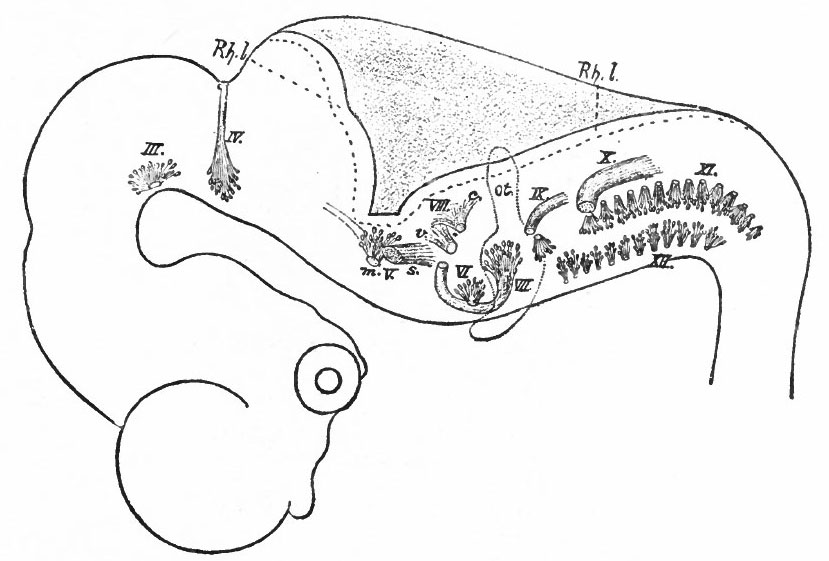
Fig. 389. Diagram (lateral view) of the brain of a 10.2 mm. human embryo (during the fifth week)
Showing the roots of the cranial nerves. His. Ill, Oculomotor; IV, Trochlear; V, Trigeminus (m, efferent root, s, afferent root) ; VI, Abducens; VII, Facial; VIII, Acoustic (c, cochlear part, v t vestibular part); IX, Glossopharyiigeus; X, Vagus; XI, Spinal accessory; XII, Hypoglossus. ot., Auditory vesicle; Rh.l., rhombic lip. The two series of efferent roots (medial and lateral) are clearly shown.
- Text-Book of Embryology: Germ cells | Maturation | Fertilization | Amphioxus | Frog | Chick | Mammalian | External body form | Connective tissues and skeletal | Vascular | Muscular | Alimentary tube and organs | Respiratory | Coelom, Diaphragm and Mesenteries | Urogenital | Integumentary | Nervous System | Special Sense | Foetal Membranes | Teratogenesis | Gallery of All Figures
| Historic Disclaimer - information about historic embryology pages |
|---|
 |
Reference
Bailey FR. and Miller AM. Text-Book of Embryology (1921) New York: William Wood and Co.
Cite this page: Hill, M.A. (2024, April 26) Embryology Bailey389.jpg. Retrieved from https://embryology.med.unsw.edu.au/embryology/index.php/File:Bailey389.jpg
- © Dr Mark Hill 2024, UNSW Embryology ISBN: 978 0 7334 2609 4 - UNSW CRICOS Provider Code No. 00098G
File history
Click on a date/time to view the file as it appeared at that time.
| Date/Time | Thumbnail | Dimensions | User | Comment | |
|---|---|---|---|---|---|
| current | 00:39, 30 January 2011 | | 829 × 561 (65 KB) | S8600021 (talk | contribs) | ==Fig. 389. Diagram (lateral view) of the brain of a 10.2 mm. human embryo (during the fifth week)== Showing the roots of the cranial nerves. His. Ill, Oculomotor; IV, Trochlear; V, Trigeminus (m, efferent root, s, afferent root) ; VI, Abducens; VII, Fac |
You cannot overwrite this file.
File usage
The following 3 pages use this file: