File:Bailey370.jpg

Original file (975 × 1,084 pixels, file size: 242 KB, MIME type: image/jpeg)
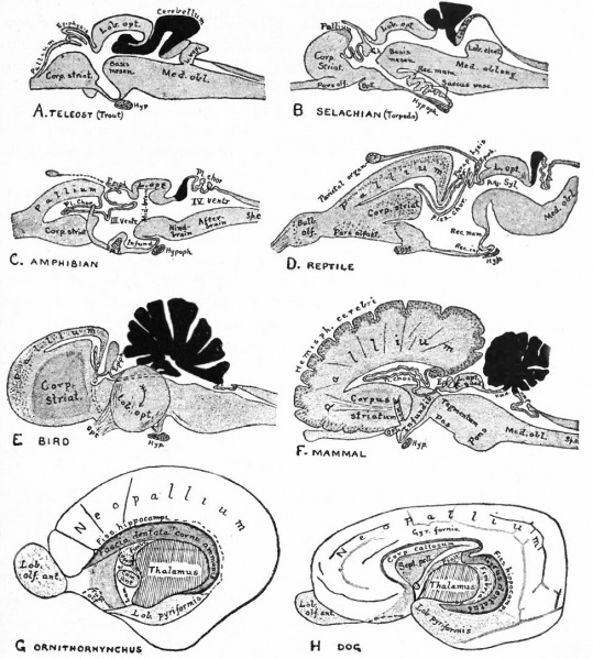
Fig. 370. Sagittal sections showing structures lying in the Median Line and Paired structures lying to one side of the median line
Sagittal sections showing structures lying in the Median Line and also paired structures (e.g., pallium) lying to one side of the median line
(Edinger)
The cerebellum is black. It is doubtful whether the membranous roof in A indicated as pallium is strictly homologous with that structure in other forms, In B, Pallium indicates prepallial structures.
Aq. SyL, Aquseductus Sylvii; Basis mesen., basis mesencephali; Bulb, olf., bulbus olfactorius; Corp. striat., corpus striatum; Epiph., epiphysis; G. h., ganglion habenulae; Hyp., hypophysis; Infund., infundibulum; Lam. t., lamina terminalis; Lob. elect., lobus electricus; L. vagi, lobus vagi; L. opt., mid-brain roof; Med. obi., medulla oblongata; Opt., optic nerve; Pl.chor., plexus chorioideus; Rec. inf., recessus infundibuli; Rec. mam., recessus mammillaris; Saccus vase., saccus vasculosus; Sp. c., spinal cord; ventr., ventricle; v. m. a., velum medullare anterius; v.m. p., velum medullare posterius.
- G and H show the mesial surface of the cerebral hemispheres in a low (G) and high (H) Mammal. G. Elliot Smith, Edinger, slightly modified.
The exposed gray matter of the olfactory regions is shaded, the darker shade indicating the archipallium (preterminal area and hippocampal formation), the lighter shade indicating the rhinencephalon, which consists of the anterior and the posterior (principally pyriform) olfactory lobes. In Amphibia and Reptiles the hippocampal formation includes all or nearly all of the mesial surface. As the early neopallium appears in the lateral hemisphere walls, the neopallial commissural fibers first pass across the median line in the ventral or anterior commissure. With the increase of the neopallium and its extension on the mesial hemisphere walls, its commissural fibers pass across more dorsally via the archipallial or fornix commissure (psalterium) forming the neopallial commissure or corpus callosum, the great development of which nearly obliterates the anterior hippocampal formation.
Com. ant., Anterior commissure; corp. callosum, corpus callosum; Fimbr., fimbria; Fiss. hippocampi, hippocampal fissure; Lam. t., lamina terminalis; Lob. olf. ant., anterior olfactory lobe; Lob. pyrfformis pyriform lobe; Psalt., psalterium (fornix commissure); Sept. pell., septum pellucidum; Tuo. olf., tuberculum olfactorium. Only a part of the gray (cortex) of the hippocampal formation appears, as the gyrus dentatus, on the mesial surface; the remainder forms an eminence, the cornu Ammonis, on the ventricular surface. This invagination is indicated extenu'lyby the hippocampal fissure. The exposed fiber bundle forming the edge of this formation (fimbria) passes forward (fornix and its commissure) and thence descends, as the anterior pillar of the fornix, behind the anterior commissure. The anterior pillar is partly indicated by a few lines in this region in the figure.
- Text-Book of Embryology: Germ cells | Maturation | Fertilization | Amphioxus | Frog | Chick | Mammalian | External body form | Connective tissues and skeletal | Vascular | Muscular | Alimentary tube and organs | Respiratory | Coelom, Diaphragm and Mesenteries | Urogenital | Integumentary | Nervous System | Special Sense | Foetal Membranes | Teratogenesis | Gallery of All Figures
| Historic Disclaimer - information about historic embryology pages |
|---|
 |
Reference
Bailey FR. and Miller AM. Text-Book of Embryology (1921) New York: William Wood and Co.
Cite this page: Hill, M.A. (2024, April 27) Embryology Bailey370.jpg. Retrieved from https://embryology.med.unsw.edu.au/embryology/index.php/File:Bailey370.jpg
- © Dr Mark Hill 2024, UNSW Embryology ISBN: 978 0 7334 2609 4 - UNSW CRICOS Provider Code No. 00098G
File history
Click on a date/time to view the file as it appeared at that time.
| Date/Time | Thumbnail | Dimensions | User | Comment | |
|---|---|---|---|---|---|
| current | 16:44, 29 January 2011 | | 975 × 1,084 (242 KB) | S8600021 (talk | contribs) | {{Template:Bailey 1921 Figures}} Category:Neural |
You cannot overwrite this file.