File:Bailey366.jpg
Original file (558 × 633 pixels, file size: 97 KB, MIME type: image/jpeg)
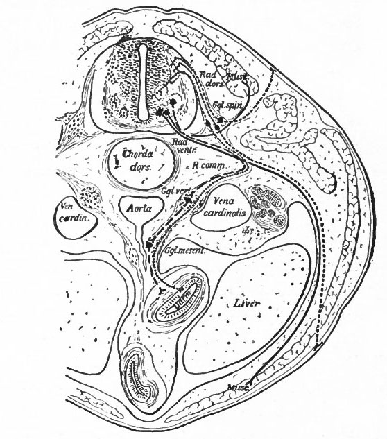
Fig. 366. Transverse section through the body of a typical Vertebrate
Showing the peripheral (segmental) nervous apparatus.
Froriep.
Small dots, afferent visceral neurones; coarse dots, afferent somatic neurones; dashes, efferent visceral (ventral root and sympathetic) neurones; lines, efferent somatic neurones.
Darm, gut; Ggl. spin., spinal ganglion; Ggl. vert., vertebral sympathetic ganglion; Ggl. mesent., mesenteric sympathetic ganglion. The peripheral sympathetic ganglionic plexuses (Auerbach and Meissner) are not shown. Muse., muscle; Rad. dors., dorsal root; Rad. vent., ventral root; R. comm., white ramus communicans.
Two sympathetic neurones are represented as intercalated in the visceral efferent pathway. It doubtful if there should be more than one.
- Text-Book of Embryology: Germ cells | Maturation | Fertilization | Amphioxus | Frog | Chick | Mammalian | External body form | Connective tissues and skeletal | Vascular | Muscular | Alimentary tube and organs | Respiratory | Coelom, Diaphragm and Mesenteries | Urogenital | Integumentary | Nervous System | Special Sense | Foetal Membranes | Teratogenesis | Gallery of All Figures
| Historic Disclaimer - information about historic embryology pages |
|---|
 |
Reference
Bailey FR. and Miller AM. Text-Book of Embryology (1921) New York: William Wood and Co.
Cite this page: Hill, M.A. (2024, May 19) Embryology Bailey366.jpg. Retrieved from https://embryology.med.unsw.edu.au/embryology/index.php/File:Bailey366.jpg
- © Dr Mark Hill 2024, UNSW Embryology ISBN: 978 0 7334 2609 4 - UNSW CRICOS Provider Code No. 00098G
File history
Click on a date/time to view the file as it appeared at that time.
| Date/Time | Thumbnail | Dimensions | User | Comment | |
|---|---|---|---|---|---|
| current | 16:41, 29 January 2011 | | 558 × 633 (97 KB) | S8600021 (talk | contribs) | ==Fig. 366. Transverse section through the body of a typical Vertebrate== Showing the peripheral (segmental) nervous apparatus. Froriep. Small dots, afferent visceral neurones; coarse dots, afferent somatic neurones; dashes, efferent visceral (ventra |
You cannot overwrite this file.