File:Bailey085.jpg
Bailey085.jpg (505 × 425 pixels, file size: 59 KB, MIME type: image/jpeg)
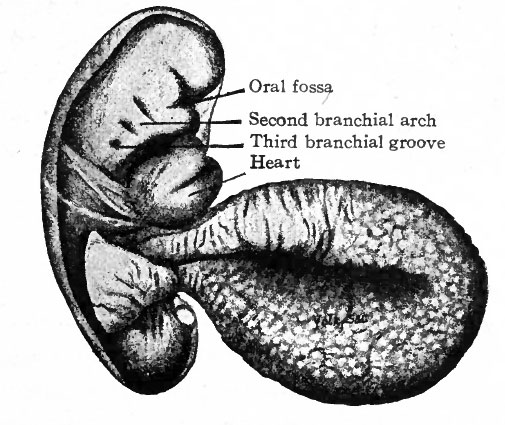
Fig. 85. Human embryo of 2.6 mm
His, from Keibel and Mall. Human Embryology
One of the early human embryos described by His is shown in Fig. 85. The veil-like structure around the embryo is the amnion. This embryo measures 2.6 mm. and was estimated to be 18-21 days old (the estimate in the light of more recent studies probably being too low). The body is more robust than in the preceding stage. In addition to the cephalic flexure the dorsum in profile is a curve, with three rather prominent regions of curvature; a cervical flexure, a dorsal flexure and a sacral flexure. The whole embryo is slightly twisted around its long axis, the head turned toward the left and the caudal end toward the right. In the cervical region are three vertical depressions which diminish in size from before backward. Alternating with these are prominences which also diminish from before backward. These alternating depressions and prominences are the branchial grooves and arches which are homologues of the gill slits and gill bars in fishes. The first arch lies in front of the first groove and bounds the oral fossa laterally; its two subdivisions, the mandibular process and maxillary process, with the notch between representing the future angle of the mouth, are already differentiated. Through the development of the first arch the depth of the oral fossa is considerably increased. The heart causes a conspicuous protrusion on the ventral side of the cervical region. The constriction between the body of the embryo and the yolk sac is marked, and this attenuated portion of the yolk sac is from now on spoken of as the yolk stalk. The structure attached caudal the yolk stalk and turned over the right side of the embryo is the belly stalk which later will be included in the umbilical cord.
- Links: Fig. 85 in text
- Text-Book of Embryology: Germ cells | Maturation | Fertilization | Amphioxus | Frog | Chick | Mammalian | External body form | Connective tissues and skeletal | Vascular | Muscular | Alimentary tube and organs | Respiratory | Coelom, Diaphragm and Mesenteries | Urogenital | Integumentary | Nervous System | Special Sense | Foetal Membranes | Teratogenesis | Gallery of All Figures
| Historic Disclaimer - information about historic embryology pages |
|---|
 |
Reference
Bailey FR. and Miller AM. Text-Book of Embryology (1921) New York: William Wood and Co.
Cite this page: Hill, M.A. (2024, April 27) Embryology Bailey085.jpg. Retrieved from https://embryology.med.unsw.edu.au/embryology/index.php/File:Bailey085.jpg
- © Dr Mark Hill 2024, UNSW Embryology ISBN: 978 0 7334 2609 4 - UNSW CRICOS Provider Code No. 00098G
File history
Click on a date/time to view the file as it appeared at that time.
| Date/Time | Thumbnail | Dimensions | User | Comment | |
|---|---|---|---|---|---|
| current | 15:36, 18 January 2011 | | 505 × 425 (59 KB) | S8600021 (talk | contribs) |
You cannot overwrite this file.
File usage
The following 3 pages use this file: