File:Anencephaly ultrasound.jpg: Difference between revisions
From Embryology
mNo edit summary |
mNo edit summary |
||
| Line 9: | Line 9: | ||
====Copyright==== | ====Copyright==== | ||
Alorainy IA, Barlas NB, Al-Boukai AA. | |||
http://creativecommons.org/licenses/by-nc-sa/3.0/ | http://creativecommons.org/licenses/by-nc-sa/3.0/ | ||
Revision as of 13:16, 22 March 2013
Anencephaly Ultrasound
Prenatal ultrasound done at 18 weeks (GA) shows coronal images of the face and orbits with symmetric and complete absence of the cranial vault and brain.
- arrows in A - large and prominent orbits.
- arrows in B - complete absence of the cranial vault and brain.
Reference
<pubmed>21042439</pubmed>| Indian J Radiol Imaging.
Copyright
Alorainy IA, Barlas NB, Al-Boukai AA.
http://creativecommons.org/licenses/by-nc-sa/3.0/
Figure 1 IndianJRadiolImaging_2010_20_3_174_69349_f4.jpg original image size adjusted.
File history
Click on a date/time to view the file as it appeared at that time.
| Date/Time | Thumbnail | Dimensions | User | Comment | |
|---|---|---|---|---|---|
| current | 13:10, 22 March 2013 | 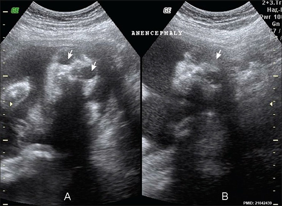 | 900 × 658 (108 KB) | Z8600021 (talk | contribs) | ==Anencephaly ultrasound== Prenatal ultrasound done at 18 weeks shows coronal images of the face and orbits with symmetric and complete absence of the cranial vault and brain. * arrows in A - large and prominent orbits. * arrows in B - complete absen... |
You cannot overwrite this file.