File:Keibel Mall 2 489.jpg
Original file (855 × 1,000 pixels, file size: 97 KB, MIME type: image/jpeg)
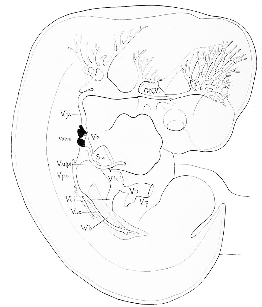
Fig. 489. Reconstruction of the right jugular lymphatic sac, shown in solid black against the jugular vein, in a human embryo 10.5 mm long
(Mall's collection, No. 109.) X about 14.
G.N.V., Gasserian ganglion; S.v., sinus venosus; V.c, vena cephalica; Y.c.i., vena cava inferior; V.h., vena hepatica; V.j.i., vena jugularis interna; V.p., vena portse; V.p.c, vena cardinalis posterior; V.sc, vena subcardinalis; V.u.(p-), vena ulnaris (primitiva); V.u., vena umbilicalis; TF.6., Wolffian body.
- IV. The Development of the Lymphatic System: Chapter XVIII. Development of Blood, Vascular System, and Spleen | Historic Disclaimer
Reference
Sabin FR. The Development of the Lymphatic System in Keibel F. and Mall FP. Manual of Human Embryology II. (1912) J. B. Lippincott Company, Philadelphia.
Cite this page: Hill, M.A. (2026, September 30) Embryology Keibel Mall 2 489.jpg. Retrieved from https://embryology.med.unsw.edu.au/embryology/index.php/File:Keibel_Mall_2_489.jpg
- © Dr Mark Hill 2026, UNSW Embryology ISBN: 978 0 7334 2609 4 - UNSW CRICOS Provider Code No. 00098G
File history
Yi efo/eka'e gwa ebo wo le nyangagi wuncin ye kamina wunga tinya nan
| Gwalagizhi | Nyangagi | Dimensions | User | Comment | |
|---|---|---|---|---|---|
| current | 14:27, 2 March 2014 | | 855 × 1,000 (97 KB) | Z8600021 (talk | contribs) |
You cannot overwrite this file.
File usage
The following page uses this file: