Category:Uterus
From Embryology
This page lists UNSW Embryology content related to development of the female uterus.
Subcategories
This category has the following 3 subcategories, out of 3 total.
Pages in category 'Uterus'
The following 72 pages are in this category, out of 72 total.
E
M
P
- Paper - Cytological studies on the internal secretory functions in the human placenta and decidua (1921)
- Paper - Development of the uterine glands in man (1920)
- Paper - Observations on the origin of the Mullerian groove in human embryos
- Paper - The chorion and endometrium of the embryo H.R.1.
- Paper - The duration of life of the spermatozoa in the human uterine tube
- Paper - The Internal Genital Organs of a Female Foetus of 15 cm Length
- Paper - Transuterine (internal) migration of the ovum in sheep and other mammals
- Template:Paramesonephric duct
- Placenta - Maternal Decidua
- Template:Placenta abnormalities
- Template:Placenta accreta
- Template:Placenta histology
- Template:Placenta increta
- Template:Placenta percreta
- Template:Placenta previa
- Template:Placenta vascular
R
- Template:Ref-AIHW-2019 Cervical Screening
- Template:Ref-BakerHookSeveringhaus1944
- Template:Ref-BaumgartnerNelsonDock1920
- Template:Ref-Boyd1944
- Template:Ref-Cohen1952
- Template:Ref-Corner1921b
- Template:Ref-Fujimura1921
- Template:Ref-Gruenwald1943a
- Template:Ref-Hart1893
- Template:Ref-Hart1896
- Template:Ref-Hart1896b
- Template:Ref-Hegar1908
- Template:Ref-Meyer1917
- Template:Ref-Minot1889
- Template:Ref-Ohli1935
- Template:Ref-Papanicolaou1933
- Template:Ref-ReynoldsBaker1951
- Template:Ref-SelyeMcKeown1934
- Template:Ref-WislockiDempsey1945
U
Media in category 'Uterus'
The following 121 files are in this category, out of 121 total.
 Bailey073.jpg 705 × 501; 122 KB
 Bailey310.jpg 688 × 365; 41 KB
 Bailey334.jpg 857 × 558; 77 KB
 Bailey335.jpg 590 × 468; 55 KB
 Bailey339.jpg 802 × 602; 111 KB
 Bailey492.jpg 907 × 695; 123 KB
 Bailey494.jpg 875 × 769; 157 KB
 Bailey498.jpg 949 × 751; 180 KB
 Bailey501.jpg 688 × 1,093; 276 KB
 Bicornuate uterus01.jpg 1,452 × 1,691; 209 KB
 Braune 1877 plate 2 fig2.jpg 806 × 1,000; 200 KB
 Braune 1877 plate 2 fig3.jpg 893 × 1,000; 208 KB
 Braune 1877 plate 29B.jpg 868 × 1,200; 322 KB
 Braune 1877 plate 30.jpg 857 × 1,200; 295 KB
 Braune 1877 plate 31.jpg 870 × 1,200; 299 KB
 Cat embryo ovary.jpg 505 × 492; 47 KB
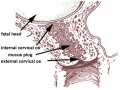 Cervical mucus plug.jpg 500 × 373; 55 KB
 Cervicovaginal mucus rheology.jpg 800 × 438; 74 KB
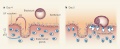
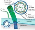 Cilium cartoon.jpg 800 × 682; 114 KB
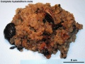 Complete hydatidiform mole 05.jpg 1,280 × 960; 324 KB
 Complete hydatidiform mole 06.jpg 1,280 × 960; 553 KB
 Corner-table02.jpg 800 × 647; 107 KB
 Corner001.jpg 858 × 356; 44 KB
 Corner1920 fig01.jpg 1,000 × 606; 159 KB
 DaviesHarding1944 fig02.jpg 617 × 800; 130 KB
 Faulconer1951 fig01.jpg 1,000 × 770; 327 KB
 Faulconer1951 fig02.jpg 1,014 × 847; 153 KB
 Faulconer1951 fig03.jpg 1,014 × 847; 206 KB
 Faulconer1951 fig04.jpg 1,014 × 847; 215 KB
 Faulconer1951 fig05.jpg 1,014 × 847; 233 KB
 Faulconer1951 fig06.jpg 1,102 × 851; 253 KB
 Faulconer1951 fig07.jpg 884 × 847; 187 KB
 Faulconer1951 plate01.jpg 2,105 × 2,746; 1.18 MB
 Female genital tract chlamydia trachomatis infection 01.jpg 804 × 500; 78 KB
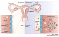
 Female reproductive tract Wnt4.jpg 1,000 × 563; 79 KB
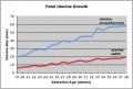 Fetal uterus growth.jpg 438 × 296; 28 KB
 Gray0033.jpg 334 × 700; 58 KB
 Gray0540.jpg 1,355 × 1,000; 261 KB
 Gray0589.jpg 900 × 534; 134 KB
 Gray0620.jpg 706 × 600; 127 KB
 Gray1109.jpg 464 × 487; 56 KB
 Gray1161.jpg 1,000 × 671; 138 KB
 Gray1166.jpg 750 × 750; 194 KB
 Gray1170.jpg 1,000 × 718; 170 KB
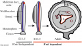
 Hamster uterus GDF8 expression.jpg 800 × 423; 57 KB
 Hertig1956 fig07.jpg 745 × 562; 104 KB
 Hertig1956 fig08.jpg 745 × 562; 77 KB
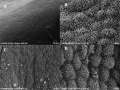
 Human - uterine epithelium SEM01.jpg 600 × 400; 37 KB
 Human - uterine epithelium SEM02.jpg 600 × 400; 26 KB
 Human - uterine epithelium TEM01.jpg 600 × 433; 25 KB
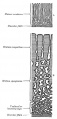
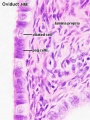
 Human fetal uterus myometrium.jpg 500 × 554; 86 KB
 Human right ovary and tube 1.jpg 916 × 680; 32 KB
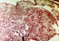
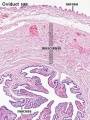
 Human uterine glands 01.jpg 726 × 545; 99 KB
 Human uterine tube ciliated epithelium SEM.jpg 1,200 × 855; 248 KB
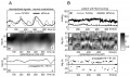 Human uterus contractile activity.jpg 1,200 × 714; 318 KB
 Human week 10 fetus 03.jpg 1,600 × 1,200; 370 KB
 Human week 10 fetus 23.jpg 1,600 × 1,200; 393 KB
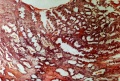
 Human- late proliferative uterine endometrium.jpg 509 × 350; 98 KB
 Human- late secretory uterine endometrium.jpg 518 × 350; 93 KB
 Human- menstrual uterine endometrium.jpg 516 × 350; 93 KB
 Human- mid-proliferative uterine endometrium.jpg 513 × 350; 111 KB
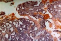
 Human- secretory uterine endometrium.jpg 515 × 350; 93 KB
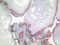
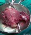
 Hydatidiform mole 01.jpg 800 × 592; 634 KB
 Hydatidiform mole 02.jpg 585 × 1,100; 237 KB
 Hydatidiform mole metastasis 01.jpg 712 × 800; 67 KB
 Implantation LIF.jpg 800 × 325; 48 KB
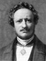 Johannes Muller.jpg 298 × 398; 20 KB
 Keibel Mall 079-082.jpg 683 × 906; 147 KB
 Keibel Mall 083-087.jpg 681 × 738; 75 KB
 Keibel Mall 094.jpg 680 × 791; 98 KB
 Keibel Mall 2 623.jpg 1,549 × 1,709; 437 KB
 Keibel Mall 2 658a.jpg 1,127 × 1,200; 103 KB
 Keibel Mall 2 658c.jpg 1,000 × 1,019; 90 KB
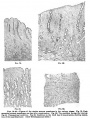
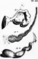
 Keith1902 fig081.jpg 818 × 800; 113 KB
 Keith1902 fig088.jpg 788 × 1,000; 92 KB
 Keith1921 fig024.jpg 945 × 957; 138 KB
 Kollmann108.jpg 800 × 767; 110 KB
 Kollmann451.jpg 796 × 826; 102 KB
 Kollmann452.jpg 687 × 606; 74 KB
 Kollmann453.jpg 664 × 566; 65 KB
 Kollmann454.jpg 729 × 641; 84 KB
 Kollmann457.jpg 777 × 598; 60 KB
 Minot1897 002.jpg 800 × 416; 134 KB
 Minot1897 003.jpg 1,176 × 708; 374 KB
 Minot1897 006.jpg 600 × 849; 168 KB
 Minot1897 007.jpg 600 × 657; 148 KB
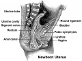 Newborn uterus.jpg 540 × 400; 72 KB
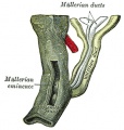
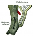
 Paramesonephric duct.jpg 423 × 478; 40 KB
 Paramesonephric ducts.jpg 371 × 400; 43 KB
 Pig - uterine epithelium SEM.jpg 800 × 598; 118 KB
 Reinier De Graaf - Plate XIX uterine tube drawings.jpg 393 × 600; 46 KB
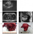
 Septate uterus ultrasound 01.jpg 1,280 × 525; 95 KB
 Skene1898 fig242.jpg 395 × 549; 62 KB
 Skene1898 fig32.jpg 500 × 414; 26 KB
 Skene1898 fig33.jpg 545 × 534; 48 KB
 Smear- early proliferative.jpg 521 × 350; 47 KB
 Smear- late secretory.jpg 525 × 350; 74 KB
 Smear- late-proliferative.jpg 511 × 350; 46 KB
 Smear- mid-proliferative.jpg 519 × 350; 58 KB
 Smear- secretory.jpg 520 × 350; 57 KB
 Smooth muscle histology 008.jpg 1,280 × 1,024; 629 KB
 Smooth muscle histology 009.jpg 1,280 × 1,024; 307 KB
 Ultrasound uterine and ovarian vascularity.jpg 531 × 780; 132 KB
 Unicornate uterus.jpg 250 × 241; 10 KB
 Uterine abnormalities.jpg 400 × 294; 15 KB
 Uterine anomalies ESHRE-ESGE classification.jpg 1,000 × 684; 93 KB
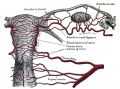
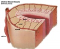 Uterine arterial vessel cartoon.jpg 600 × 483; 44 KB
 Uterine gland proliferative phase.jpg 400 × 533; 51 KB
 Uterine gland secretory phase.jpg 400 × 533; 49 KB
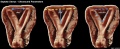
 Uterine teratoma.jpg 600 × 609; 73 KB
 Uterine tube histology 02.jpg 400 × 533; 55 KB
 Uterine tube histology 03.jpg 400 × 533; 34 KB
 Uterine tube histology 04.jpg 1,280 × 1,024; 253 KB
 Uterine tube histology 05.jpg 1,063 × 1,063; 508 KB
 Uterine tube histology.jpg 1,280 × 1,024; 568 KB
 Uterus anatomical position.jpg 500 × 373; 62 KB
 Uterus proliferative phase.jpg 400 × 533; 52 KB
 Uterus secretory phase 01.jpg 1,280 × 1,024; 318 KB
 Uterus secretory phase 02.jpg 1,280 × 1,024; 359 KB
 Uterus secretory phase.jpg 400 × 533; 68 KB
 Week1 summary.jpg 1,000 × 747; 107 KB


