Category:Morula: Difference between revisions
From Embryology
No edit summary |
No edit summary |
||
| Line 4: | Line 4: | ||
'''Links:''' [[Week 1]] | [[Fertilization]] | '''Links:''' [[Week 1]] | [[Fertilization]] | ||
[[Category:Week 1]] | [[Category:Week 1]] | ||
Revision as of 14:34, 11 November 2011
The pages and media below are UNSW Embryology content that relates to morula development.
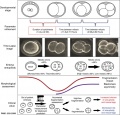
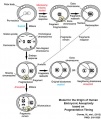
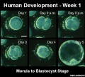
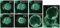
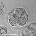
(Latin, morula = mulberry) An early stage in post-fertilization development when cells have rapidly divided to produce a solid mass of cells (12-15 cells) with a "mulberry" appearance. This stage is followed by formation of a cavity in this cellular mass (blastocyst stage). In humans, morula stage of development occurs during the first week following fertilization.
Links: Week 1 | Fertilization
Subcategories
This category has the following 3 subcategories, out of 3 total.
Pages in category 'Morula'
The following 18 pages are in this category, out of 18 total.
M
P
Media in category 'Morula'
The following 50 files are in this category, out of 50 total.
 Aneuploidy model based on fragmentation 1.jpg 946 × 907; 154 KB
 Aneuploidy model based on fragmentation.jpg 668 × 790; 105 KB
 Bailey053.jpg 769 × 195; 27 KB
 Bailey080.jpg 766 × 738; 120 KB
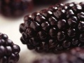 Blackberry.jpg 392 × 294; 25 KB
 Blastomere isolation.jpg 1,200 × 961; 95 KB
 Blastomere mitotic spindle orientation.jpg 600 × 414; 21 KB
 Bovine morula 01.jpg 800 × 802; 84 KB
 Bovine morula and blastocyst 01.jpg 989 × 1,200; 231 KB
 Canine oocyte to blastocyst.jpg 825 × 1,000; 154 KB
 Early human telomere length.jpg 1,800 × 1,034; 70 KB
 Early human telomeres.jpg 1,280 × 1,006; 194 KB
 Early mouse development cartoon.jpg 1,000 × 430; 81 KB
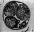 Hamilton1945-fig12.jpg 594 × 550; 59 KB
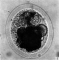 Hamilton1945-fig13.jpg 974 × 1,000; 150 KB
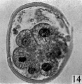 Hamilton1945-fig14.jpg 536 × 548; 55 KB
 Hamilton1945-fig15.jpg 576 × 548; 61 KB
 Hamilton1945-plate03.jpg 1,507 × 2,000; 347 KB
 Human blastocyst day 1-5.jpg 500 × 450; 46 KB
 Human blastocyst day 1-6.jpg 708 × 338; 45 KB
 Human blastocyst day 3-6.mov ; 4.26 MB
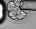
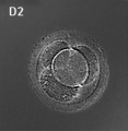 Human embryo day 2.jpg 400 × 409; 19 KB
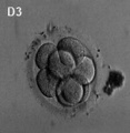 Human embryo day 3.jpg 400 × 409; 7 KB
 Human preimplantation embryos 01.jpg 1,280 × 629; 261 KB
 Keith1921 fig012.jpg 1,164 × 595; 137 KB
 Model embryo to 32 cell stage 001.jpg 696 × 256; 22 KB
 Model embryo to 32 cell stage 240.jpg 696 × 256; 49 KB
 Model embryo to 32 cell stage icon.jpg 696 × 256; 30 KB
 Model human blastocyst development.jpg 946 × 726; 84 KB
 Mouse - blastocoel formation.jpg 800 × 319; 47 KB
 Mouse blastocyst movie icon.jpg 480 × 480; 26 KB
 Mouse blastocyst movie pronuclei.jpg 480 × 480; 32 KB
 Mouse blastocyst movie.mp4 ; 860 KB
 Mouse inner cell mass cell types 01.jpg 820 × 800; 128 KB
 Mouse Sox2 expression 01.jpg 1,000 × 375; 63 KB
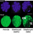 Mouse Sox2 expression 03.jpg 600 × 615; 55 KB
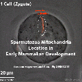
 Mouse spermatozoa mito movie icon.jpg 495 × 495; 43 KB
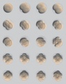
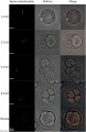 Mouse spermatozoa mitochondria 01.jpg 831 × 1,280; 141 KB
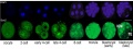
 Mouse- preimplantation gene expression 01.jpg 726 × 1,959; 226 KB
 Mouse- preimplantation gene expression 02.jpg 657 × 659; 74 KB
 Mouse- preimplantation gene expression 03.jpg 633 × 510; 61 KB
 Mouse- preimplantation gene expression 04.jpg 714 × 660; 80 KB
 Mouse- preimplantation gene expression 05.jpg 622 × 363; 30 KB
 Mouse- preimplantation gene expression.jpg 800 × 612; 106 KB
 Mouse-embryo granzyme G.jpg 899 × 1,000; 90 KB
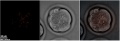
 Mouse-morula 01.jpg 400 × 398; 15 KB
 Preimplantation blastomere biopsy.jpg 626 × 500; 75 KB
 Sea Urchin- early embryo cleavage pattern.jpg 600 × 761; 153 KB
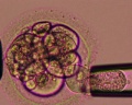
 Spermatozoa mitochondria morula.jpg 906 × 306; 38 KB
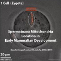
 Spermatozoa mitochondria PMID23878233.gif 495 × 495; 974 KB


