Category:Limb: Difference between revisions
From Embryology
mNo edit summary |
mNo edit summary |
||
| Line 1: | Line 1: | ||
This page lists {{Embryology}} content related to limb development and abnormalities. The limb is also an excellent model for how tissue pattern is established. | This page lists {{Embryology}} content related to limb development and abnormalities. The limb is also an excellent model for how tissue pattern is established. | ||
:'''Links:''' [[Musculoskeletal System - Limb Development|Limb Development]] | [[Book_-_Manual_of_Human_Embryology_11D| | |||
:'''Links:''' [[Musculoskeletal System - Limb Development|Limb Development]] | [[Musculoskeletal System - Limb Abnormalities|Limb Abnormalities]] | [[Musculoskeletal System - Appendicular Skeleton Development|Appendicular Skeleton]] | [[Book_-_Manual_of_Human_Embryology_11D|1910 - Limb Development]] | |||
Revision as of 11:16, 11 October 2014
This page lists Embryology content related to limb development and abnormalities. The limb is also an excellent model for how tissue pattern is established.
Pages in category 'Limb'
The following 148 pages are in this category, out of 148 total.
B
- Template:Bardeen1906 figures
- BGDA Practical 7 - Week 6
- Book - An Atlas of Topographical Anatomy 19
- Book - An Atlas of Topographical Anatomy 20
- Book - An Atlas of Topographical Anatomy 21
- Book - An Atlas of Topographical Anatomy 22
- Book - An Atlas of Topographical Anatomy 23
- Book - An Atlas of Topographical Anatomy 24
- Book - An Atlas of Topographical Anatomy 26
- Book - An Atlas of Topographical Anatomy 27
- Book - An Atlas of Topographical Anatomy 28
- Book - Human Embryology and Morphology 20
- Book - Manual of Human Embryology 11D
- Book - Manual of Human Embryology 12
C
D
L
- Lecture - Limb Development
- Template:Limb
- Template:Limb abnormalities
- Template:Limb axis
- Template:Limb Links
- Template:Limb Rotation table
- Template:Limb skeleton mesenchyme-cartilage-bone table
- Template:Lower limb ossification collapse table
- Template:Lower limb ossification collapsible table
- Template:Lower limb ossification table
- Template:Lower limb ossification timeline
M
P
- Paper - A contribution to the embryology of the fore-limb
- Paper - A study of the development of the mammalian pelvis
- Paper - An unusual form of brachyphalangy and syndactyly with double proximal phalanx in the middle fingers (1932)
- Paper - Anatomy of a seven months foetus exhibiting bilateral absence of the ulna accompanied by monodactyly
- Paper - Chondrification in the hands and feet of staged human embryos
- Paper - Development and variation of the nerves and the musculature of the inferior extremity and of the neighboring regions of the trunk in man
- Paper - Development of the innervation pattern in the upper limb of staged human embryos (1990)
- Paper - Extra digits and digital reductions
- Paper - Familial abnormalities of the middle phalanges of each hand (1923)
- Paper - On an instance of two subclavian arteries of the early arm bud of man and its fundamental significance
- Paper - Rare congenital malformation of hands and feet (1924)
- Paper - Studies of the development of the human skeleton (1905)
- Paper - The arrangement of the bursae in the superior extremities of the full-time foetus
- Paper - The development and evolution of the "papillary" ridges and patterns on the volar surface of the hand (1906)
- Paper - The development of joints
- Paper - The development of the arm in man (1902)
- Paper - The development of the arteries of the human lower extremity
- Paper - The development of the limbs, body-wall and back
- Paper - The development of the limbs, body-wall and back (1901)
- Paper - The development of the patella
- Paper - The development of the principal arterial stems in the forelimb of the pig (1922)
- Paper - The development of the veins in the limbs of rabbit embryos
- Paper - The early development of the knee joint in staged human embryos
- Paper - The embryology of the human hip joint (1943)
- Paper - The epiphysis of the head of the femur (1915)
- Paper - The growth of the human foot
- Paper - The proximo-distal sequence of origin of the parts of the chick wing and the role of the ectoderm
- Paper - The sexual differences of the fetal pelvis
- Paper - The sexual differences of the fetal pelvis (1899)
- Template:Pelvis
- Template:Pelvis Timeline table
R
- Template:Ref-Bardeen1907
- Template:Ref-BardeenLewis1901
- Template:Ref-Brailsford1943
- Template:Ref-Carey1922
- Template:Ref-Cockayne1932
- Template:Ref-Cummins1929
- Template:Ref-Davenport1932
- Template:Ref-deLima1928
- Template:Ref-Evans1908
- Template:Ref-Evatt1906
- Template:Ref-GardnerO'Rahilly1968
- Template:Ref-Geddes1912b
- Template:Ref-GrayGardener1950
- Template:Ref-Haines1947
- Template:Ref-Haines1953
- Template:Ref-Hann1923
- Template:Ref-Harrison1915
- Template:Ref-Johnson1899
- Template:Ref-Keith1910
- Template:Ref-Lewis1902
- Template:Ref-Lewis1905b
- Template:Ref-Lewis1910a
- Template:Ref-Manson1924
- Template:Ref-O'Rahilly1957
- Template:Ref-PMID55906
- Template:Ref-Prentiss1906
- Template:Ref-Rutherford1914
- Template:Ref-Saunders1947a
- Template:Ref-Saunders1948
- Template:Ref-ShinoharaTanaka1990
- Template:Ref-Stacey1938
- Template:Ref-Walmsley1915
- Template:Ref-Walmsley1940
- Template:Ref-Watt1917
- Template:Ref-Whittaker1910
- Template:Ref-Wigoder1928
- Template:Ref-Woollard1922
S
U
V
Media in category 'Limb'
The following 74 files are in this category, out of 274 total.
(previous page) (next page)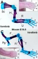 Mouse limb cartilage and bone E18.5.jpg 774 × 1,200; 152 KB
 Mouse limb gene expression 01.mov ; 1.12 MB
 Mouse limb gene expression 01.mp4 ; 1.34 MB
- Mouse limb gene expression 02.mp4 ; 1.34 MB
 Mouse limb gene expression icon.jpg 446 × 550; 48 KB
 Mouse limb skeleton cartoon.jpg 1,000 × 487; 64 KB
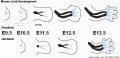
 Mouse limb tissue development.jpg 1,280 × 767; 161 KB
 Mouse Lmx1b gene expression.jpg 1,624 × 550; 104 KB
 Mouse pax7 limb 01.jpg 1,320 × 549; 112 KB
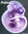 Mouse Prdm1.jpg 446 × 550; 35 KB
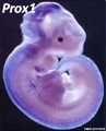 Mouse Prox1.jpg 446 × 550; 36 KB
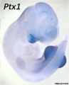 Mouse Ptx1.jpg 446 × 550; 21 KB
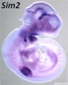 Mouse Sim2.jpg 446 × 550; 30 KB
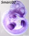 Mouse SmarcD2.jpg 446 × 550; 32 KB
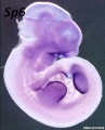 Mouse Sp6.jpg 446 × 550; 34 KB
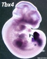 Mouse Tbx4.jpg 446 × 550; 37 KB
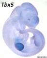 Mouse Tbx5.jpg 446 × 550; 21 KB
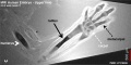
 Mouse- forelimb bud Shh expression and BMP pathway.jpg 905 × 308; 35 KB
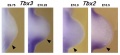 Mouse- forelimb-bud-Tbx3-Tbx2.jpg 955 × 436; 45 KB
 Mouse- Hand2 forelimb buds.jpg 458 × 600; 48 KB
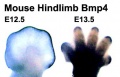 Mouse- hindlimb Bmp4 expression.jpg 374 × 241; 19 KB
 Mouse- hindlimb buds Shh expression.jpg 600 × 679; 30 KB
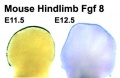 Mouse- hindlimb Fgf8 expression.jpg 374 × 243; 17 KB
 Mouse-E17.5 foot.jpg 385 × 500; 29 KB
 Mouse-E17.5 normal and abnormal limb.jpg 661 × 848; 105 KB
 Mouse-E17.5-foot-large.jpg 600 × 779; 53 KB
 Mouse-forelimb bud Fgf4-Hoxd13-Hoxa13.jpg 881 × 237; 30 KB
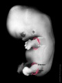
 MRI Human Embryo - upper limb 01.jpg 1,418 × 940; 106 KB
 MRI Human Embryo - upper limb 02.jpg 1,000 × 497; 74 KB
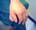
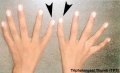
 Nail patella syndrome 02.jpg 809 × 525; 51 KB
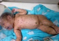 Neonatal varicella.jpg 795 × 561; 35 KB
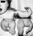
 Polydactylia.jpg 700 × 541; 37 KB
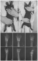
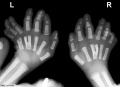 Polydactyly and y-shaped central metacarpals x-ray.jpg 800 × 579; 51 KB
 Polydactyly.jpg 230 × 188; 10 KB
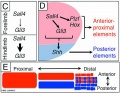
 Posterior forelimb bud identity.jpg 600 × 406; 46 KB
 Posterior forelimb bud identity.png 600 × 406; 895 KB
 Rabbitmalformation3.jpg 363 × 574; 44 KB
 Sall4 limb skeletal elements model.jpg 800 × 619; 88 KB
 Senior1919 fig10.jpg 1,280 × 679; 134 KB
 Shoulder cartoon.jpg 592 × 597; 36 KB
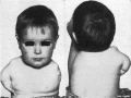
 Sirenomelia 01.jpg 488 × 600; 23 KB
 Sirenomelia 02.jpg 498 × 598; 14 KB
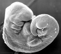
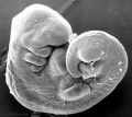
 Stage13 sem1.jpg 1,000 × 886; 80 KB
 Stage13 sem1a.jpg 800 × 709; 57 KB
 Stage13 sem1b.jpg 600 × 532; 36 KB
 Stage13 sem1c.jpg 400 × 355; 18 KB
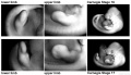 Stage16-17-limbs01.jpg 1,000 × 572; 77 KB
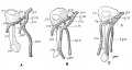
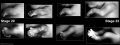
 Stage19- limb rotation.jpg 600 × 800; 27 KB
 Stage20-23 limbs a.jpg 800 × 301; 19 KB
 Stage20-23 limbs b.jpg 600 × 226; 13 KB
 Stage20-23 limbs.jpg 1,000 × 376; 26 KB
 Streeter1920chart3.jpg 1,200 × 635; 79 KB
 Streeter1920table4.jpg 1,120 × 593; 80 KB
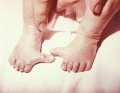
 Syndactyly.jpg 443 × 377; 16 KB
 Syndactyly2.JPG 220 × 221; 7 KB
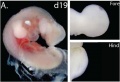 Tammar wallaby limb day 19 development.jpg 700 × 477; 39 KB
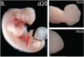 Tammar wallaby limb day 20 development.jpg 700 × 477; 36 KB
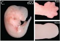 Tammar wallaby limb day 22 development.jpg 700 × 477; 36 KB
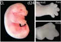 Tammar wallaby limb day 24 development.jpg 700 × 477; 39 KB
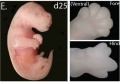 Tammar wallaby limb day 25 development.jpg 700 × 477; 37 KB
 Tammar wallaby limb development 01.jpg 1,200 × 551; 101 KB
 Thalidomide - CPS49 vascular effect.jpg 1,000 × 1,002; 197 KB
 Thalidomide - limb signaling.jpg 628 × 540; 88 KB
 Thalidomide abnormalities 01.jpg 800 × 598; 71 KB
 Thalidomide abnormalities 02.jpg 724 × 800; 74 KB
 Thomson1899 fig03.jpg 1,209 × 1,678; 82 KB
 Triphalangeal-thumb.jpg 450 × 273; 14 KB
 Woollard-plate01.jpg 744 × 1,000; 153 KB
 Woollard-plate02.jpg 788 × 1,000; 196 KB
 Woollard001.jpg 739 × 858; 142 KB
 Woollard002.jpg 699 × 873; 125 KB
 Woollard003.jpg 1,107 × 848; 159 KB
 Woollard004.jpg 1,037 × 717; 148 KB
 Woollard005.jpg 1,034 × 655; 144 KB


