Category:Inner Ear
From Embryology
This Embryology category shows pages and media related to the inner ear development.
- Links: inner ear
Pages in category 'Inner Ear'
The following 53 pages are in this category, out of 53 total.
A
H
P
- Paper - 1880 The Platypus Cochlea
- Paper - 1906 Observations on the Labyrinth of Certain Animals
- Paper - 1917 The Typical Form of the Cochlea and Its Variations
- Paper - Development of the aquaductus cochleae and the periotic (perilymphatic) duct
- Paper - On the development of the membrana tectoria with reference to its structure and attachments
- Paper - On the proportions, development and attachment of the tectorial membrane (1915)
- Paper - The cytological processes involved in the formation of the scalae of the internal ear
- Paper - The development and structure of the otic (endolymphatic) sac
- Paper - The development of the cochlear fenestra, fossula and secondary tympanic membrane
- Paper - The development of the otic capsule in the region of the vestibular aqueduct
- Paper - The Development of the Scala Tympani, Scala Vestibuli and Perioticular Cistern in the Human Embryo
- Paper - The developmental course of the human auditory vesicle
- Paper - The early development of the otic vesicle in staged human embryos
- Paper - The Factors Involved in the Excavation of the Cavities in the Cartilaginous Capsule of the Ear in the Human Embryo
- Paper - The vascular drainage of the endolymphatic sac and its topographical relation to the transverse sinus in the human
- Template:Placode
- Template:Placodes
R
- Template:Ref-AnsonBast1951a
- Template:Ref-AnsonBast1951b
- Template:Ref-AnsonBast1958b
- Template:Ref-Ayers1919
- Template:Ref-Hardesty1915a
- Template:Ref-Hardesty1915b
- Template:Ref-Keen1939
- Template:Ref-Keen1940
- Template:Ref-Prentiss1913
- Template:Ref-Pritchard1880
- Template:Ref-Streeter1917innerear3
- Template:Ref-Stricht1919
- Template:Ref-Stricht1919b
S
Media in category 'Inner Ear'
The following 81 files are in this category, out of 81 total.
 Adult cochlea cartoon 01.jpg 986 × 800; 123 KB
 Adult cochlea nerve glia cartoon.jpg 1,000 × 725; 85 KB
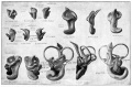
 Anson1934 fig01-8.jpg 1,337 × 888; 126 KB
 Anson1934 fig09.jpg 546 × 272; 16 KB
 Anson1934 fig10.jpg 520 × 612; 31 KB
 Anson1934 fig11.jpg 758 × 870; 52 KB
 Anson1934 fig12.jpg 545 × 968; 44 KB
 Anson1934 fig13.jpg 761 × 1,323; 74 KB
 Anson1934 fig14.jpg 761 × 1,323; 81 KB
 Anson1934 fig15.jpg 693 × 1,003; 62 KB
 Anson1934 fig16.jpg 691 × 985; 47 KB
 Anson1934 fig17.jpg 605 × 1,143; 57 KB
 Anson1934 fig18.jpg 418 × 1,161; 34 KB
 Anson1934 fig19.jpg 524 × 1,218; 48 KB
 Anson1934 plate01.jpg 1,557 × 2,279; 288 KB
 Anson1934 plate02.jpg 1,464 × 2,311; 259 KB
 Bailey474.jpg 1,321 × 869; 247 KB
 Cat inner ear MicroCT.jpg 1,159 × 1,300; 266 KB
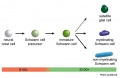 Cochlea glial lineage cartoon.jpg 1,000 × 651; 52 KB
 Cochlea stria vascularis cartoon 01.jpg 874 × 1,897; 338 KB
 Cochlea stria vascularis cartoon 02.jpg 802 × 800; 138 KB
 Cochlea stria vascularis cartoon 03.jpg 694 × 800; 125 KB
 Gray0903.jpg 600 × 389; 42 KB
 Human cochlea fetal development cartoon.jpg 592 × 1,200; 96 KB
 Human cochlea stria vascularis 01.jpg 1,854 × 1,806; 754 KB
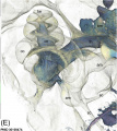
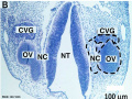 Human CS13 otic vesicle 01.jpg 1,028 × 774; 112 KB
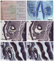 Human CS13-15 otic vesicle 01.jpg 1,574 × 1,779; 364 KB
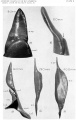
 Human fetal cochlea 01.jpg 1,270 × 532; 266 KB
 Human fetal cochlea 02.jpg 1,270 × 532; 271 KB
 Human inner ear MicroCT.jpg 2,131 × 3,111; 1,001 KB
 Inner ear development cartoon 01.jpg 714 × 800; 94 KB
 Inner ear haircells.jpg 800 × 671; 121 KB
 Lewis1906 fig438.jpg 1,007 × 877; 149 KB
 Macklin1914 fig08.jpg 1,790 × 1,004; 170 KB
 Macklin1914 fig09.jpg 1,685 × 1,045; 180 KB
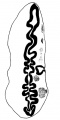
 Max Brödel-cochlea drawing1934.jpg 531 × 395; 69 KB
 Minot1897 429.jpg 1,200 × 805; 189 KB
 Mouse cochlea development cartoon.jpg 1,000 × 280; 53 KB
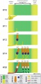
 Mouse cochlea gene expression.jpg 1,000 × 346; 75 KB
 Mouse organ of corti 01.jpg 1,280 × 1,024; 339 KB
 Mouse organ of corti 02.jpg 1,280 × 1,024; 320 KB
 Mouse organ of corti 03.jpg 1,280 × 1,024; 207 KB
 Mouse organ of corti 04.jpg 1,280 × 1,024; 202 KB
 Mouse organ of corti 05.jpg 1,280 × 1,024; 171 KB
 Mouse organ of corti NeuroD1.jpg 1,779 × 2,383; 1.11 MB
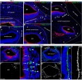
 Mouse otic placode gene expression 01.jpg 358 × 677; 86 KB
 Mouse otic placode gene expression 02.jpg 500 × 486; 100 KB
 Neural domain.jpg 452 × 778; 54 KB
 Stage 22 image 218.jpg 1,200 × 730; 308 KB
 Streeter026.jpg 774 × 1,000; 45 KB
 Streeter027.jpg 774 × 1,000; 51 KB
 Streeter028-30.jpg 748 × 1,000; 134 KB
 Streeter028.jpg 774 × 1,000; 69 KB
 Streeter029.jpg 774 × 1,000; 74 KB
 Streeter030.jpg 774 × 1,000; 78 KB
 Streeter031.jpg 774 × 1,000; 79 KB
 Streeter1906 fig04.jpg 2,237 × 1,113; 404 KB
 Streeter1906 plate01.jpg 2,708 × 1,786; 701 KB
 Streeter1906 plate02.jpg 2,783 × 1,819; 499 KB
 Streeter1917-fig01.jpg 1,128 × 800; 298 KB
 Streeter1917-fig02.jpg 1,000 × 847; 276 KB
 Streeter1917-fig03.jpg 1,000 × 420; 173 KB
 Streeter1917-fig04-05.jpg 1,000 × 544; 124 KB
 Streeter1917-fig04.jpg 536 × 544; 61 KB
 Streeter1917-fig05.jpg 556 × 544; 64 KB
 Streeter1917-fig06-07.jpg 1,337 × 1,671; 639 KB
 Streeter1917-fig06.jpg 539 × 749; 93 KB
 Streeter1917-fig07.jpg 584 × 749; 105 KB
 Streeter1917-fig08-09.jpg 1,200 × 853; 218 KB
 Streeter1917-fig08.jpg 610 × 853; 110 KB
 Streeter1917-fig09.jpg 617 × 853; 109 KB
 Streeter1957 fig07.jpg 1,280 × 1,201; 117 KB
 Streeter1957 fig7-19.jpg 680 × 800; 48 KB
 Streeter1957 fig7-20.jpg 680 × 800; 24 KB
 Streeter1957 fig7-21.jpg 680 × 800; 25 KB
 Streeter1957 fig7-22.jpg 680 × 800; 33 KB
 Streeter1957 fig7-23.jpg 680 × 800; 37 KB
 Stricht1919b plate1.jpg 1,280 × 1,844; 352 KB
 Stricht1919b plate2.jpg 1,280 × 1,938; 311 KB
 Stricht1919b plate3.jpg 1,280 × 1,960; 348 KB
 West09.jpg 403 × 806; 44 KB


