Category:Human
From Embryology
This Embryology category shows pages and media related to human development.
Subcategories
This category has the following 72 subcategories, out of 72 total.
C
- Carnegie Embryo
- Carnegie Embryo 1
- Carnegie Embryo 112
- Carnegie Embryo 1134B
- Carnegie Embryo 116
- Carnegie Embryo 1266
- Carnegie Embryo 1455
- Carnegie Embryo 148
- Carnegie Embryo 172
- Carnegie Embryo 19
- Carnegie Embryo 239
- Carnegie Embryo 2393
- Carnegie Embryo 240
- Carnegie Embryo 248
- Carnegie Embryo 256
- Carnegie Embryo 296
- Carnegie Embryo 3527
- Carnegie Embryo 3956
- Carnegie Embryo 4046
- Carnegie Embryo 4059
- Carnegie Embryo 407
- Carnegie Embryo 4148
- Carnegie Embryo 43
- Carnegie Embryo 4405
- Carnegie Embryo 460
- Carnegie Embryo 463
- Carnegie Embryo 523
- Carnegie Embryo 5541
- Carnegie Embryo 5609
- Carnegie Embryo 5652
- Carnegie Embryo 5682
- Carnegie Embryo 5874
- Carnegie Embryo 6032
- Carnegie Embryo 625
- Carnegie Embryo 6426
- Carnegie Embryo 6581
- Carnegie Embryo 7618
- Carnegie Embryo 7669
- Carnegie Embryo 786
- Carnegie Embryo 808
- Carnegie Embryo 8147
- Carnegie Embryo 8239
- Carnegie Embryo 8370
- Carnegie Embryo 858
- Carnegie Embryo 8630
- Carnegie Embryo 8967
- Carnegie Embryo 9296
- Carnegie Embryo 96
- Carnegie Embryo 963
- Carnegie Embryo 966
- Carnegie Embryo 9697
D
F
H
Pages in category 'Human'
The following 124 pages are in this category, out of 332 total.
(previous page) (next page)P
- Paper - Models of the pancreas in embryos of the pig, rabbit, cat, and man (1908)
- Paper - Notes on the development of the human sphenoid (1910)
- Paper - Notes on the postnatal growth of the heart kidneys liver and spleen in man (1919)
- Paper - Observations on the neural crest of a ten-somite human embryo (1939)
- Paper - On The Age Of Human Embryos
- Paper - On the Development of the Human Heart
- Paper - On the Development, Ossification, and Growth of the Palate Bone of Man
- Paper - On the Frequency of Localized Anomalies in Human Embryos and Infants at Birth
- Paper - Phases of Maturation and Fertilization in Human Ova
- Paper - Significant superficial anastomoses in the arterial blood supply to the human brain (1959)
- Paper - Some Gross Structural and Quantitative Aspects of the Developmental Anatomy of the Human Embryonic, Fetal and Circumnatal Skeleton
- Paper - Some Observations on the Development of the Ventral Pancreas in Man
- Paper - Teratogenecity in the setting of cardiac development and maldevelopment
- Paper - Teratological studies (1919)
- Paper - The Anatomy of Human Embryos with Seventeen to Twenty-three Pairs of Somites
- Paper - The cartilaginous skull of a human embryo twenty-one millimeters in length (1920)
- Paper - The cortex of the brain in the human embryo during the fourth month with special reference to the so-called Papilla of Retzius
- Paper - The critical period in the development of the intestines (1914)
- Paper - The development and reduction of the tail and of the caudal end of the spinal cord (1920)
- Paper - The Development of Head-Process and Prochordal Plate in Man
- Paper - The development of synovial joints
- Paper - The Development of the Cranial and Spinal Nerves in the Occipital Region of the Human Embryo
- Paper - The development of the human prostate gland with reference to the development of other structures at the neck of the urinary bladder (1912)
- Paper - The development of the mucous membrane oesophagus stomach and small intestine in human embryo
- Paper - The development of the nervous tissues of the human embryo (1877)
- Paper - The Development of the Nose and of the Pharynx and its Derivatives in Man
- Paper - The Development of the Scala Tympani, Scala Vestibuli and Perioticular Cistern in the Human Embryo
- Paper - The development of the subcutaneous vascular plexus in the head of the human embryo (1923)
- Paper - The developmental alterations in the vascular system of the brain of the human embryo (1921)
- Paper - The Earliest Blood-Vessels in Man
- Paper - The Early Development of Man, with Special Reference to the Development of the Mesoderm and Cloacal Membrane
- Paper - The early development of the otic vesicle in staged human embryos
- Paper - The early relation of the auditory vesicle to the ectoderm in human embryos
- Paper - The Factors Involved in the Excavation of the Cavities in the Cartilaginous Capsule of the Ear in the Human Embryo
- Paper - The first appearance of the neural tube and optic primordium in the human embryo at stage 10
- Paper - The formation of the umbilical cord and the umbilical region of the anterior abdominal wall
- Paper - The genesis and structure of the membrana tectoria and the crista spiralis of the cochlea (1918)
- Paper - The Internal Genital Organs of a Female Foetus of 15 cm Length
- Paper - The Maturation of the Human Ovum
- Paper - The Origin of the Otic and Optic Primordia in Man
- Paper - The Peripheral Nervous System in the Human Embryo at the End of the First Month (10 mm)
- Paper - The prenatal development of the human temporomandibular joint
- Paper - The relations of the somites of the head to the brain in a human embryo of 20 paired somites
- Paper - The sexual differences of the fetal pelvis
- Paper - The sexual differences of the fetal pelvis (1899)
- Paper - The subdivisions of the neural folds in man
- Paper - The vascular drainage of the endolymphatic sac and its topographical relation to the transverse sinus in the human
- Paper - Transformation of the aortic-arch system during the development of the human embryo (1922)
- Paper - Two Early Human Embryos
- Paper - Two presomite human embryos
- Paper - Vertebral Regional Determination in Young Human Embryos
- Paper The development of the subcutaneous vascular plexus in the head of the human embryo (1923)
- Patent Ductus Venosus Movie
- Template:Placental villi timeline
- Template:PMID22437671 links
- Pregnancy Test
R
- Template:Ref-BascomOsterud1925
- Template:Ref-Baumgartner1917
- Template:Ref-Berry1900
- Template:Ref-Bolk1915
- Template:Ref-Carey1919
- Template:Ref-ClarkGJ1900
- Template:Ref-Gladstone1935
- Template:Ref-HertigAdams1967
- Template:Ref-Hines1921
- Template:Ref-Jirasek1980
- Template:Ref-Johnston1914
- Template:Ref-Noback1943 figures
- Template:Ref-OtisBrent1954
- Template:Ref-Romanes1941b
- Template:Ref-Schmidt1877
- Template:Ref-Schultz1919
- Template:Ref-Siddiqi1934
- Template:Ref-Valentin1834
- Template:Ref-Veit1918
S
- Template:Second trimester timeline
- SH Lecture - Lymphatic Structure and Organs
- Template:Shoulder timeline
- Template:Shoulder Timeline table2
- Template:SlideCircumvallatePlacenta
- Template:SlideOvaryCorpusLuteum
- Template:SlidePlacentalVilli1
- Template:Smell timeline
- Template:Spaulding1922
- Template:Species Placenta collapsetable1
- Template:Species Placenta table1
- Template:Spleen timeline
- Template:Spleen Timeline Table
- Template talk:Spleen Timeline Table
- Template:Stage 11 BF images
- Template:Stage19 bf2 links
- Template:Stage23oralcavity images
- Template:Streeter1908 figures
- Template:Streeter1917
- Template:Streeter1917 figures
- Template:Streeter1917a
- Template:Streeter1919 figures
- Template:Streeter1921
- Template:Sudler1902 figures
T
U
V
W
Media in category 'Human'
The following 200 files are in this category, out of 2,421 total.
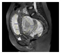
(previous page) (next page)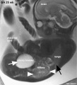 Fetal kidney MRI 01.jpg 797 × 880; 68 KB
 Fetal kidney MRI 02.jpg 797 × 880; 64 KB
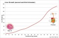
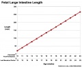 Fetal large Intestine length growth graph.jpg 800 × 653; 50 KB
 Fetal length change.jpg 972 × 648; 72 KB
 Fetal liver erythroblasts 01.jpg 905 × 534; 69 KB
 Fetal liver weight growth graph.jpg 800 × 521; 34 KB
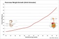 Fetal pancreas weight growth graph.jpg 1,000 × 669; 49 KB
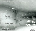 Fetal pineal gland 01.jpg 700 × 603; 67 KB
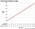 Fetal small Intestine length growth graph.jpg 800 × 653; 51 KB
 Fetal temporomandibular joint 01.jpg 600 × 391; 107 KB
 Fetal temporomandibular joint 02.jpg 600 × 390; 109 KB
 Fetal temporomandibular joint 03.jpg 600 × 393; 48 KB
 Fetal temporomandibular joint 04.jpg 600 × 390; 67 KB
 Fetal temporomandibular joint 05.jpg 600 × 392; 71 KB
 Fetal temporomandibular joint 06.jpg 600 × 389; 50 KB
 Fetal thymus.jpg 450 × 600; 122 KB
 Fetal ultrasound ductal arch 01.jpg 800 × 533; 27 KB
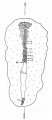
 Finley1923 fig01.jpg 494 × 968; 53 KB
 Finley1923 fig02.jpg 700 × 800; 77 KB
 Finley1923 fig03.jpg 600 × 512; 56 KB
 Finley1923 fig04.jpg 700 × 627; 61 KB
 Finley1923 fig05.jpg 674 × 800; 146 KB
 Finley1923 fig06.jpg 729 × 800; 95 KB
 Finley1923 fig07.jpg 314 × 800; 40 KB
 Finley1923 fig08.jpg 296 × 800; 32 KB
 Finley1923 fig09.jpg 456 × 800; 49 KB
 Finley1923 fig10.jpg 221 × 802; 17 KB
 Finley1923 fig11.jpg 432 × 800; 38 KB
 Finley1923 fig12.jpg 593 × 800; 48 KB
 Finley1923 fig13.jpg 594 × 800; 51 KB
 Finley1923 Plate 1.jpg 776 × 1,000; 151 KB
 Finley1923 Plate 2.jpg 864 × 1,200; 153 KB
 Fleming1927-fig01.jpg 953 × 1,000; 167 KB
 Fleming1927-fig02.jpg 1,000 × 685; 53 KB
 Fleming1927-fig03.jpg 1,200 × 1,908; 602 KB
 Fleming1927-fig03a.jpg 1,179 × 493; 150 KB
 Fleming1927-fig03b.jpg 1,179 × 577; 171 KB
 Fleming1927-fig03c.jpg 1,179 × 800; 266 KB
 Fleming1927-fig04.jpg 800 × 899; 103 KB
 Florian1933 fig01.jpg 532 × 605; 38 KB
 Florian1933 fig02.jpg 580 × 569; 37 KB
 Florian1933 fig03.jpg 602 × 593; 40 KB
 Florian1933 fig04.jpg 754 × 610; 50 KB
 Florian1933 fig05.jpg 682 × 605; 51 KB
 Florian1933 fig06.jpg 731 × 584; 55 KB
 Florian1933 fig07.jpg 1,059 × 590; 60 KB
 Florian1933 fig08.jpg 1,109 × 520; 55 KB
 Foster109.jpg 873 × 372; 70 KB
 Foster110.jpg 924 × 821; 113 KB
 Foster112.jpg 1,002 × 494; 76 KB
 Foster113.jpg 927 × 512; 57 KB
 Foster114.jpg 725 × 1,071; 135 KB
 Foster117.jpg 797 × 707; 97 KB
 Foster118b.jpg 809 × 1,077; 232 KB
 Foster122.jpg 608 × 419; 45 KB
 Foster134.jpg 342 × 503; 37 KB
 Foster138.jpg 755 × 540; 51 KB
 Frazer1926 fig01.jpg 1,200 × 804; 137 KB
 Frazer1926 fig02.jpg 991 × 833; 145 KB
 Frazer1926 fig03.jpg 1,200 × 804; 69 KB
 Frazer1926 fig04.jpg 1,200 × 804; 95 KB
 Frazer1926 fig05.jpg 1,000 × 643; 85 KB
 Frazer1926 fig06.jpg 563 × 811; 35 KB
 Frazer1926 fig07.gif 554 × 600; 134 KB
 Frazer1926 fig07.jpg 1,229 × 996; 95 KB
 Frazer1926 fig08.jpg 616 × 789; 38 KB
 Frazer1926 plate01.jpg 1,914 × 2,681; 469 KB
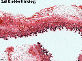
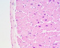
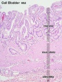 Gall bladder histology 001.jpg 375 × 500; 78 KB
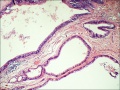
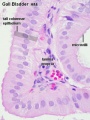 Gall bladder histology 002.jpg 375 × 500; 45 KB
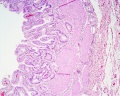 Gall bladder histology 003.jpg 1,280 × 1,024; 577 KB
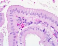 Gall bladder histology 004.jpg 1,280 × 1,024; 254 KB
 Gall bladder histology 005.gif 600 × 450; 683 KB
 Germ cell tumor 02.jpg 800 × 599; 168 KB
 Gillilan1959-fig01.jpg 1,000 × 1,313; 155 KB
 Gillilan1959-fig02.jpg 905 × 1,000; 159 KB
 Gillilan1959-fig03.jpg 1,042 × 1,366; 317 KB
 Gray0054.jpg 800 × 513; 71 KB
 Gray0070.jpg 800 × 796; 182 KB
 Gray0071.jpg 700 × 440; 104 KB
 Gray0178.jpg 617 × 368; 44 KB
 Gray0179.jpg 617 × 368; 47 KB
 Gray0180.jpg 617 × 368; 48 KB
 Gray0181.jpg 617 × 368; 52 KB
 Gray0649.jpg 698 × 700; 72 KB
 Gray0651.jpg 698 × 700; 94 KB
 Gray0652.jpg 698 × 700; 103 KB
 Gray0653.jpg 698 × 700; 108 KB
 Gray0654.jpg 402 × 500; 39 KB
 Gray0655.jpg 500 × 419; 39 KB
 Gray0658.jpg 361 × 450; 29 KB
 Gray0847.jpg 559 × 900; 155 KB
 Gray0848.jpg 800 × 935; 289 KB
 Gray0849.jpg 800 × 885; 258 KB
 Gray0865.jpg 759 × 400; 70 KB
 Gray0867.jpg 495 × 600; 131 KB
 Gray0869.jpg 748 × 600; 126 KB
 Gray0870.jpg 637 × 600; 109 KB
 Gray0871.jpg 450 × 682; 114 KB
 Gray0872.jpg 499 × 600; 93 KB
 Gray0873.jpg 919 × 545; 124 KB
 Gray0874.jpg 600 × 545; 120 KB
 Gray0875.jpg 600 × 537; 83 KB
 Gray0876.jpg 256 × 700; 75 KB
 Gray0877.jpg 500 × 794; 101 KB
 Gray0878.jpg 580 × 550; 120 KB
 Gray0879.jpg 600 × 522; 66 KB
 Gray0880.jpg 800 × 496; 147 KB
 Gray0881.jpg 800 × 536; 87 KB
 Gray0882.jpg 800 × 649; 123 KB
 Gray0883.jpg 616 × 700; 107 KB
 Gray0884.jpg 419 × 400; 58 KB
 Gray0885.jpg 667 × 400; 20 KB
 Gray0886.jpg 500 × 368; 17 KB
 Gray0887.jpg 221 × 800; 67 KB
 Gray0888.jpg 711 × 566; 110 KB
 Gray0889.jpg 800 × 519; 103 KB
 Gray0890.jpg 700 × 700; 104 KB
 Gray0891.jpg 600 × 544; 104 KB
 Gray0892.jpg 500 × 409; 47 KB
 Gray0893.jpg 355 × 700; 93 KB
 Gray0894.jpg 710 × 400; 74 KB
 Gray0895.jpg 700 × 452; 94 KB
 Gray0896.jpg 600 × 638; 68 KB
 Gray0897.jpg 500 × 366; 47 KB
 Gray0904.jpg 226 × 350; 26 KB
 Gray0905.jpg 430 × 275; 19 KB
 Gray0906.jpg 438 × 600; 81 KB
 Gray0907.jpg 679 × 600; 110 KB
 Gray0909.jpg 600 × 437; 56 KB
 Gray0910.jpg 400 × 548; 78 KB
 Gray0911.jpg 651 × 400; 73 KB
 Gray0912.jpg 600 × 540; 87 KB
 Gray0913.jpg 671 × 600; 98 KB
 Gray0914.jpg 708 × 500; 102 KB
 Gray0915.jpg 720 × 600; 94 KB
 Gray0940.jpg 618 × 700; 160 KB
 Gray0941.jpg 700 × 524; 106 KB
 Gray0942.jpg 800 × 515; 127 KB
 Gray0971.jpg 800 × 583; 166 KB
 Gray0978.jpg 483 × 600; 67 KB
 Gray0979.jpg 500 × 446; 56 KB
 Gray0980.jpg 542 × 450; 57 KB
 Gray0981.jpg 538 × 340; 50 KB
 Gray0986.jpg 565 × 606; 56 KB
 Gray1108.jpg 590 × 400; 73 KB
 Gray1109.jpg 464 × 487; 56 KB
 Gray1111.jpg 523 × 600; 66 KB
 Gray1113.jpg 600 × 385; 68 KB
 Gray1114.jpg 450 × 471; 47 KB
 Gray1115.jpg 600 × 474; 73 KB
 Gray1116.jpg 600 × 433; 88 KB
 Gray1117.jpg 581 × 510; 75 KB
 Gray1118.jpg 600 × 403; 45 KB
 Gray1119.jpg 700 × 807; 115 KB
 Gray1174.jpg 782 × 800; 165 KB
 Gray1175.jpg 500 × 500; 31 KB
 Gray1183.jpg 800 × 325; 45 KB
 Gray1184.jpg 800 × 325; 39 KB
 Gray1235.jpg 800 × 389; 41 KB
 Gray1236.jpg 800 × 281; 29 KB
 Gray1237.jpg 371 × 600; 57 KB
 Greater-omentum.jpg 537 × 419; 48 KB
 Haemomonochorial human placenta EM01.jpg 792 × 775; 79 KB
 Hamilton1944-fig01.jpg 732 × 800; 168 KB
 Hamilton1944-fig02.jpg 540 × 556; 74 KB
 Hamilton1944-fig03.jpg 732 × 800; 169 KB
 Hamilton1944-fig04.jpg 714 × 794; 189 KB
 Hamilton1944-fig05.jpg 795 × 782; 101 KB
 Hamilton1944-fig06.jpg 385 × 749; 48 KB
 Hamilton1944-fig07.jpg 873 × 800; 140 KB
 Hamilton1944-plate01.jpg 1,200 × 1,472; 387 KB
 Hamilton1944-plate02.jpg 1,200 × 1,663; 287 KB
 Hamilton1944-table01.jpg 1,200 × 645; 113 KB
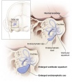 Hearing-vestibular sac abnormality.jpg 432 × 493; 45 KB
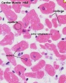
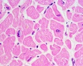
 Heart histology 001.jpg 400 × 500; 83 KB
 Heart histology 002.jpg 400 × 500; 81 KB
 Heart histology 003.jpg 400 × 500; 136 KB
 Heart histology 004.jpg 400 × 500; 97 KB
 Heart histology 101.jpg 1,280 × 1,024; 258 KB
 Heart histology 102.jpg 1,280 × 1,024; 242 KB
 Heart histology 103.jpg 1,280 × 1,024; 281 KB
 Heart histology 104.jpg 1,280 × 1,024; 280 KB
 Heart histology 105.jpg 1,280 × 1,024; 379 KB
 Heart histology 106.jpg 1,280 × 1,024; 347 KB
 Heart histology 107.jpg 1,280 × 1,024; 395 KB
 Heart human embryo CRL10mm 01.jpg 1,000 × 1,389; 537 KB
 Heart innervation 01.jpg 1,280 × 599; 92 KB
 Heart valve histology 01.jpg 1,008 × 1,280; 318 KB
 Heart valve histology 02.jpg 800 × 456; 103 KB
 Heart valve histology 03.jpg 800 × 489; 111 KB
 Heart-cartoon-001.jpg 600 × 697; 40 KB
 Heart-histology-102.jpg 1,280 × 1,024; 242 KB
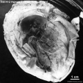
 Herring1908b fig06.jpg 1,280 × 1,234; 283 KB
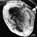
 Herring1908b fig07.jpg 1,280 × 974; 290 KB
 Heterotopic pregnancy MRI 01.jpg 600 × 536; 106 KB
 HillFlorian1931a fig01.jpg 594 × 1,372; 74 KB
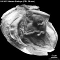 HillH12 Stage 23 bf01.jpg 1,500 × 1,500; 227 KB
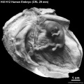 HillH12 Stage 23 bf02.jpg 1,500 × 1,500; 219 KB
 HillH12 Stage 23 bf03.jpg 1,500 × 1,500; 275 KB
 HillH12 Stage 23 bf04.jpg 1,500 × 1,500; 316 KB


