Category:Genital
From Embryology
This Embryology category shows pages and media related to development of the genital system. Note that links below beginning with "Paper -" are generally historic research articles.
Subcategories
This category has the following 26 subcategories, out of 26 total.
A
C
D
F
M
T
U
V
Pages in category 'Genital'
The following 200 pages are in this category, out of 430 total.
(previous page) (next page)C
D
E
G
- Template:Genital
- Genital - Female Development
- Genital - Male Development
- Template:Genital abnormalities
- Genital Abnormality - Hypospadias
- Template:Genital cartoons
- Template:Genital Links
- Genital Quiz
- Genital System - Abnormalities
- Talk:Genital System - Abnormalities
- Genital System Development
- Template:Genital terms
- Template:Germinal epithelium
- Template:Gonad
- Gonad Blood Supply Development
- Template:Graafian
- Template:Graafian follicle
- Template:Granulosa lutein cells
- Template:Gray1110 links
- Template:Gubernaculum
H
I
L
M
O
P
- Paper - A contribution to the development of the prostate in man (1909)
- Paper - A morphological study of testicular descent
- Paper - A note on a case of bifid penis (1924)
- Paper - A note on the origin and histogenesis of the mesonephric duct in mammals
- Paper - A previllous human embryo (1946)
- Paper - A report on two cases of hermaphroditism in man (1923)
- Paper - A study of the function of the epididymis 1 (1929)
- Paper - A suggestion as to the cause of the aspermatic condition of the imperfectly descended testis (1922)
- Paper - Adult human ovaries with follicles containing several oocytes (1912)
- Paper - An experimental study of the morphogenesis of intersexuality (1940)
- Paper - Anatomy, pathology and development of the hymen
- Paper - Anomalies of the genito-urinary tract
- Paper - Another case of hermaphroditism in man (1924)
- Paper - Chiefly concerning the genito-mesenteric fold of peritoneum
- Paper - Cysts of the genital ducts Müllerian and Wolffian (1946)
- Paper - Cytology of the human spermatozoon
- Paper - Development and transition of the testis, normal and abnormal 1
- Paper - Development and transition of the testis, normal and abnormal 2
- Paper - Development and transition of the testis, normal and abnormal 3
- Paper - Development and transition of the testis, normal and abnormal 4
- Paper - Development and vascularization of the testis (1906)
- Paper - Development of the egg of the cow up to the stage of blastocyst formation (1946)
- Paper - Development of the human ovary from birth to sexual maturity
- Paper - Development of the mammary gland
- Paper - Development of the mammary gland - Arris and Gale Lecture
- Paper - Development of the Mouse Gonads 1
- Paper - Development of the Mouse Gonads 2
- Paper - Development of the Mouse Gonads 3
- Paper - Development of the Mouse Gonads 4
- Paper - Development of the trigone of the bladder and the termination of the mesonephric ducts (1946)
- Paper - Development of the urogenital system in the Marsupialia 2
- Paper - Development of the uterine glands in man (1920)
- Paper - Development of the vagina in the human fetus
- Paper - Electron microscopy of the sperm tail - results obtained with a new fixative
- Paper - Experimental evidence regarding the role of the anterior pituitary in the development and regulation of the genital system
- Paper - Growth of the reproductive and endocrine organs of the guinea-pig (1936)
- Paper - Has a persistence of the Müllerian ducts any relation to the conditions of cryptorchidism?
- Paper - Hermaphroditism in a mole with male external genitals (1924)
- Paper - Hermaphroditism in man (1920)
- Paper - Histochemical observations on the germ cells of human embryos
- Paper - Histochemical observations on the germ cells of human embryos (1953)
- Paper - History of the development of the human ovum (1834)
- Paper - Human ova from large follicles - including a search for maturation divisions and observations on atresia
- Paper - Human ova from large follicles - including a search for maturation divisions and observations on atresia (1930)
- Paper - Hydatiform degeneration in an early human embryo (1946)
- Paper - Malformations of the human body from a new point of view 3+4
- Paper - Malformations of the human body from a new point of view 5+6
- Paper - Morphology of the human urinogenital tract (1901)
- Paper - Morphology of the tubules of the human testis and epididymis
- Paper - Note on a case of bifid penis with penial hypospadia (1914)
- Paper - Notes on the development of the prepuce (1935)
- Paper - Observations on the origin of the Mullerian groove in human embryos
- Paper - On the anlage of the bulbo-urethral and major vestibular glands in the human embryo (1915)
- Paper - On the number of chromosomes and the type of sex chromosomes in man (1934)
- Paper - On the origin and phylogenetic significance of the female genital passages
- Paper - On the phenomena of sex-differentiation (1892)
- Paper - On the postnatal development of the ovary (albino rat), with especial reference to the number of ova (1920)
- Paper - On the role of the developing epidermis in forming sheaths and lumina to organs
- Paper - Origin and early history of the primordial germ-cells in the chick (1914)
- Paper - Origin of the sex cells in man
- Paper - Origin of the sex-cords and definitive spermatogonia in the male chick (1916)
- Paper - Origin, development and degeneration of the blood vessels of the ovary (1899)
- Paper - Post-natal growth changes in the human prostate
- Paper - Preliminary note on the development of the clitoris, vagina and hymen
- Paper - Regnier De Graaf 1641-1673
- Paper - Selective elimination of ova in the adult ovary
- Paper - Selective elimination of ova in the adult ovary (1925)
- Paper - Sex-determination and sex-differentiation in mammals (1917)
- Paper - Sexual differences of the hypophyses and their determination by the gonads
- Paper - Some observations on the development of the vagina in the pig (1934)
- Paper - Some points in the nomenclature of the external genitalia of the female
- Paper - Studies in mammalian spermatogenesis II. The spermatogenesis of man
- Paper - Studies on the fine structure of the mammalian testis 1
- Paper - Studies on the mammary gland 2 (1917)
- Paper - Testes descent 1909 - 1
- Paper - Testes descent 1909 - 2
- Paper - Testes descent 1909 - 3
- Paper - The Accessory Chromosome-Sex Determinant? (1902)
- Paper - The course of the Wolffian tubules in mammalian embryos
- Paper - The development of the cloaca in human embryos
- Paper - The development of the gonads in man with a consideration of the role of fetal endocrines and the histogenesis of ovarian tumors
- Paper - The development of the human prostate gland with reference to the development of other structures at the neck of the urinary bladder (1912)
- Paper - The development of the human vagina
- Paper - The development of the hymen
- Paper - The Development of the Infra-Umbilical Portion of the Abdominal Wall, with Remarks on the Aetiology of Ectopia Vesicae
- Paper - The development of the lower end of the vagina (1927)
- Paper - The development of the penile urethra and the homology of cowper's gland of male spermophile (1937)
- Paper - The development of the prostate gland in the human female and homologies of the urethra and vagina of the sexes
- Paper - The development of the seminal vesicles in man
- Paper - The development of the sex cords in the gonads of man and mammals
- Paper - The development of the urogenital system in Marsupialia, with special reference to Trichosurus vulpecula 1
- Paper - The development of the urogenital system in Marsupialia, with special reference to Trichosurus vulpecula 2
- Paper - The development of the vagina in the rabbit (1933)
- Paper - The early development of the corpus luteum in the mare (1946)
- Paper - The Embryonic Development of the Interstitial Cells of Leydig (1904)
- Paper - The embryonic development of the ovary and testis of the mammals (1904)
- Paper - The histology of an hermaphrodite pig and its developmental significance (1929)
- Paper - The histology of the retained testis in the human subject at different ages, and its comparison with the scrotal testis (1929)
- Paper - The inguinal canal in the foetus and new-born (1944)
- Paper - The Internal Genital Organs of a Female Foetus of 15 cm Length
- Paper - The interstitial cells of the mammalian ovary (1914)
- Paper - The lower ends of the wolffian ducts in a female pig embryo (1914)
- Paper - The Maturation of the Human Ovum
- Paper - The morphogenesis of the mammalian ovary (1913)
- Paper - The morphology of the external genitalia of the mammala (1914)
- Paper - The morphology of the seminiferous tubules of Mammalia (1913)
- Paper - The mucinous changes of the vaginal epithelium of certain mammals in pregnancy (1915)
- Paper - The nature of the malformations of the rectum and urogenital passages
- Paper - The occurrence of polyovular graafian follicles (1924)
- Paper - The origin of the lutein cells of the corpus luteum
- Paper - The physiological descent of the ovaries in the human foetus
- Paper - The relation of the growing Mullerian duct to the Wolffian duct and its importance for the genesis of malformations
- Paper - The ripe human Graafian follicle, together with some suggestions as to its mode of rupture
- Paper - The Terminal Part of the Wolffian Duct
- Paper - Transverse septal atresia of the lower third of the genital tract
Media in category 'Genital'
The following 200 files are in this category, out of 510 total.
(previous page) (next page) Allen1904 plate7.jpg 1,280 × 1,837; 704 KB
 Alternate androgen synthesis pathway.jpg 750 × 425; 99 KB
 Anogenital distance from birth to 2 years.jpg 565 × 545; 34 KB
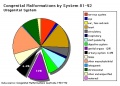 Australian abnormalities 81-92 urogenital.jpg 600 × 429; 54 KB
 Azoospermia.jpg 768 × 554; 77 KB
 Bacterial vaginosis.png 1,000 × 755; 134 KB
 Bailey307.jpg 832 × 833; 128 KB
 Bailey308.jpg 791 × 848; 88 KB
 Bailey309.jpg 594 × 592; 58 KB
 Bailey310.jpg 688 × 365; 41 KB
 Bailey327.jpg 872 × 567; 89 KB
 Bailey328.jpg 704 × 795; 85 KB
 Bailey329.jpg 838 × 381; 70 KB
 Bailey330.jpg 680 × 539; 109 KB
 Bailey331.jpg 890 × 782; 118 KB
 Bailey332.jpg 637 × 356; 53 KB
 Bailey333.jpg 830 × 445; 74 KB
 Bailey334.jpg 857 × 558; 77 KB
 Bailey335.jpg 590 × 468; 55 KB
 Bailey336.jpg 912 × 597; 145 KB
 Bailey337.jpg 790 × 573; 74 KB
 Bailey338.jpg 940 × 473; 87 KB
 Bailey339.jpg 802 × 602; 111 KB
 Bailey341.jpg 832 × 675; 69 KB
 Bailey342.jpg 777 × 561; 55 KB
 Bailey343-344.jpg 1,185 × 630; 91 KB
 Bailey345-346.jpg 1,193 × 491; 79 KB
 Bailey347-348.jpg 1,198 × 526; 83 KB
 Baileytable06.jpg 800 × 258; 27 KB
 Baileytable07.jpg 960 × 833; 152 KB
 BGDB2011-Embryo Lab 010611-01.mp3 ; 3.46 MB
- BGDB2011-Embryo Lab 010611-02.mp3 ; 2.99 MB
- BGDB2011-Embryo Lab 010611-03.mp3 ; 2.66 MB
- BGDB2011-Embryo Lab 010611-04.mp3 ; 2.94 MB
- BGDB2011-Embryo Lab 010611-05.mp3 ; 5.17 MB
 Braune 1877 plate 29B.jpg 868 × 1,200; 322 KB
 Braune 1877 plate 30.jpg 857 × 1,200; 295 KB
 Braune 1877 plate 31.jpg 870 × 1,200; 299 KB
 Bulmer1957 plate01.jpg 1,280 × 1,808; 353 KB
 Cat embryo ovary.jpg 505 × 492; 47 KB
 Caudal duplication syndrome.jpg 700 × 599; 47 KB
 Chicken primordial germ cell migration model.jpg 750 × 447; 69 KB
 Corner1920 fig01.jpg 1,000 × 606; 159 KB
 Corner1920 fig02-05.jpg 1,000 × 671; 216 KB
 Corner1920 Plate 1.jpg 756 × 1,000; 222 KB
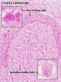 Corpus luteum lutein cells.jpg 450 × 600; 104 KB
 Corpus luteum.jpg 450 × 600; 94 KB
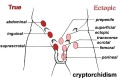 Cryptorchidism.jpg 600 × 390; 35 KB
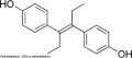 Diethylstilbestrol.jpg 600 × 263; 16 KB
 Dog- spermatozoa NANOG expression.jpg 800 × 691; 109 KB
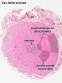 Ductus deferens 01.jpg 400 × 533; 76 KB
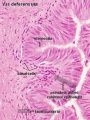 Ductus deferens 02.jpg 400 × 533; 80 KB
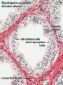
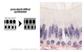 Epididymis histology 01.jpg 600 × 375; 20 KB
 Epididymis histology 02.jpg 400 × 534; 71 KB
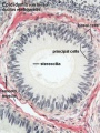 Epididymis histology 03.jpg 400 × 533; 68 KB
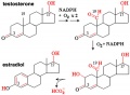 Estradiol synthesis.jpg 600 × 441; 40 KB
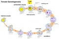 Female gametogenesis.jpg 1,000 × 666; 94 KB
 Female genital and ureter abnormality 01.jpg 766 × 732; 86 KB
 Female genital and ureter abnormality 02.jpg 766 × 733; 78 KB
 Female genital and ureter abnormality 03.jpg 766 × 762; 79 KB
 Female genital tract chlamydia trachomatis infection 01.jpg 804 × 500; 78 KB
 Female reproductive tract Wnt4.jpg 1,000 × 563; 79 KB
 Female- OHVIRA syndrome 01.jpg 340 × 1,000; 76 KB
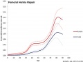 Femoral hernia repair.jpg 800 × 596; 52 KB
 Fetal 10wk urogenital 1.jpg 800 × 600; 109 KB
 Fetal 10wk urogenital 2.jpg 800 × 600; 110 KB
 Fetal 10wk urogenital 3.jpg 800 × 600; 107 KB
 Fetal 10wk urogenital 4.jpg 800 × 600; 105 KB
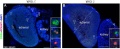
 Fetal adrenal ectopic germ cells 01.jpg 1,092 × 1,280; 358 KB
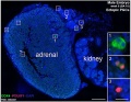
 Fetal adrenal ectopic germ cells 02.jpg 1,086 × 446; 124 KB
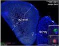
 Fetal adrenal ectopic germ cells 03.jpg 899 × 700; 159 KB
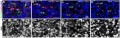
 Fetal adrenal ectopic germ cells 04.jpg 899 × 700; 147 KB
 Fetal ovary meiosis 01.jpg 1,280 × 410; 132 KB
 Fetal ovary meiosis 02.jpg 496 × 600; 77 KB
 Fetal ovary meiosis 03.jpg 652 × 400; 64 KB
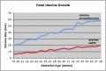 Fetal uterus growth.jpg 438 × 296; 28 KB
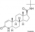 Finasteride.jpg 500 × 463; 18 KB
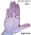 Finger length ratio - 2D4D.jpg 428 × 480; 26 KB
 Fleming1927-fig01.jpg 953 × 1,000; 167 KB
 Fleming1927-fig02.jpg 1,000 × 685; 53 KB
 Fleming1927-fig03.jpg 1,200 × 1,908; 602 KB
 Fleming1927-fig03a.jpg 1,179 × 493; 150 KB
 Fleming1927-fig03b.jpg 1,179 × 577; 171 KB
 Fleming1927-fig03c.jpg 1,179 × 800; 266 KB
 Fleming1927-fig04.jpg 800 × 899; 103 KB
 Fragile X protein cartoon.jpg 1,000 × 615; 107 KB
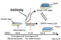 Gadd45g and sex determination model.jpg 909 × 600; 62 KB
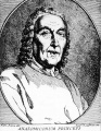 Giovanni Battista Morgagni.jpg 778 × 1,000; 183 KB
 Gonad blood 01 icon.jpg 296 × 413; 28 KB
 Gray0594.jpg 600 × 432; 83 KB
 Gray0619.jpg 800 × 741; 180 KB
 Gray0847.jpg 559 × 900; 155 KB
 Gray0849.jpg 800 × 885; 258 KB
 Gray0982a.jpg 427 × 393; 18 KB
 Gray0982b.jpg 427 × 393; 20 KB
 Gray0986.jpg 565 × 606; 56 KB
 Gray0991.jpg 469 × 400; 33 KB
 Gray0992.jpg 600 × 611; 73 KB
 Gray1108.jpg 590 × 400; 73 KB
 Gray1109.jpg 464 × 487; 56 KB
 Gray1110.jpg 414 × 1,200; 86 KB
 Gray1110a.jpg 578 × 600; 40 KB
 Gray1110b.jpg 578 × 600; 44 KB
 Gray1110c.jpg 578 × 600; 49 KB
 Gray1111.jpg 523 × 600; 66 KB
 Gray1112.jpg 550 × 548; 51 KB
 Gray1113.jpg 600 × 385; 68 KB
 Gray1114.jpg 450 × 471; 47 KB
 Gray1115.jpg 600 × 474; 73 KB
 Gray1116.jpg 600 × 433; 88 KB
 Gray1117.jpg 581 × 510; 75 KB
 Gray1118.jpg 600 × 403; 45 KB
 Gray1119.jpg 700 × 807; 115 KB
 Gray1137.jpg 600 × 502; 64 KB
 Gray1138.jpg 656 × 472; 57 KB
 Gray1139.jpg 600 × 524; 73 KB
 Gray1152.jpg 597 × 600; 100 KB
 Gray1153.jpg 725 × 600; 100 KB
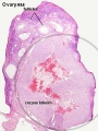
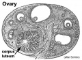 Historic-ovary.jpg 385 × 283; 34 KB
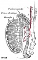 Historic-testis.jpg 509 × 800; 67 KB
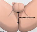 Human anogenital distance.jpg 570 × 499; 23 KB
 Human Embryology Manual 2 19 - Urogenital fold table.jpg 1,280 × 816; 166 KB
 Human idiogram-chromosome X.jpg 31 × 293; 3 KB
 Human infant ovary follicle 01.jpg 800 × 800; 107 KB
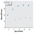
 Human male anogenital distance graph.jpg 600 × 429; 28 KB
 Human ovary postnatal growth.jpg 800 × 467; 40 KB
 Human X chromosome.jpg 400 × 450; 25 KB
 Human Y chromosome 01.jpg 937 × 549; 167 KB
 Human Y chromosome SRY region.jpg 351 × 517; 17 KB
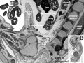
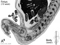 Human- fetal week 10 lower body A.jpg 600 × 450; 96 KB
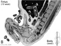 Human- fetal week 10 lower body B.jpg 600 × 450; 93 KB
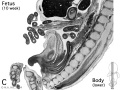 Human- fetal week 10 lower body C.jpg 600 × 450; 94 KB
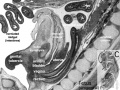
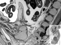
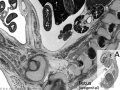 Human- fetal week 10 urogenital A.jpg 600 × 450; 109 KB
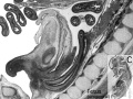
 Human- fetal week 10 urogenital B.jpg 600 × 450; 109 KB
 Human- fetal week 10 urogenital C.jpg 600 × 450; 105 KB
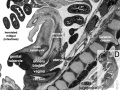
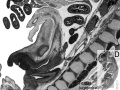 Human- fetal week 10 urogenital D.jpg 600 × 450; 101 KB
 Human- genital development critical periods.jpg 1,000 × 494; 78 KB
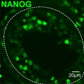 Human- spermatozoa NANOG expression 01.jpg 798 × 797; 79 KB
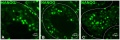 Human- spermatozoa NANOG expression.jpg 1,000 × 333; 77 KB
 Hydrocolpos.jpg 375 × 361; 23 KB
 Hypospadia 3D ultrasound 01.jpg 1,150 × 497; 87 KB
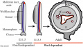
 Hypospadia classifications.jpg 500 × 410; 47 KB
 Infant ovary.jpg 943 × 571; 108 KB
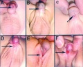
 Inguinal hernia repair 2.jpg 800 × 718; 53 KB
 Inguinal hernia repair.jpg 800 × 597; 46 KB
 Keibel Mall 2 552.jpg 1,280 × 948; 257 KB
 Keibel Mall 2 553.jpg 1,280 × 1,376; 496 KB
 Keibel Mall 2 554a.jpg 1,280 × 933; 221 KB
 Keibel Mall 2 554b.jpg 1,280 × 798; 153 KB
 Keibel Mall 2 554c.jpg 1,280 × 872; 188 KB
 Keibel Mall 2 556.jpg 1,280 × 1,473; 312 KB
 Keibel Mall 2 557.jpg 1,000 × 665; 167 KB
 Keibel Mall 2 558.jpg 1,280 × 792; 132 KB
 Keibel Mall 2 626.jpg 850 × 679; 143 KB
 Keibel Mall 2 627.jpg 1,000 × 1,252; 169 KB
 Keibel Mall 2 628.jpg 1,000 × 512; 65 KB
 Keibel Mall 2 632.jpg 1,000 × 684; 100 KB
 Keibel Mall 2 635.jpg 1,000 × 1,073; 223 KB
 Keibel Mall 2 636.jpg 1,200 × 738; 119 KB
 Keibel Mall 2 641.jpg 1,041 × 518; 62 KB
 Keibel Mall 2 642.jpg 1,200 × 644; 95 KB
 Keibel Mall 2 643.jpg 975 × 1,000; 95 KB
 Keibel Mall 2 645.jpg 957 × 1,000; 98 KB
 Keibel Mall 2 646.jpg 1,280 × 1,307; 181 KB
 Keibel Mall 2 652.jpg 1,200 × 1,036; 130 KB
 Keibel Mall 2 656.jpg 1,200 × 746; 152 KB
 Keibel Mall 2 658a.jpg 1,127 × 1,200; 103 KB
 Keibel Mall 2 658b.jpg 895 × 1,200; 98 KB
 Keibel Mall 2 658c.jpg 1,000 × 1,019; 90 KB
 Keith1902 fig079.jpg 742 × 800; 78 KB
 Keith1902 fig080.jpg 651 × 700; 79 KB
 Keith1902 fig081.jpg 818 × 800; 113 KB
 Keith1902 fig082.jpg 924 × 800; 98 KB
 Keith1902 fig083.jpg 782 × 700; 79 KB
 Keith1902 fig084.jpg 732 × 800; 88 KB
 Keith1902 fig085.jpg 800 × 590; 78 KB
 Keith1902 fig086.jpg 842 × 700; 84 KB
 Keith1902 fig087.jpg 800 × 613; 90 KB
 Keith1902 fig088.jpg 788 × 1,000; 92 KB
 Keith1902 fig089.jpg 964 × 800; 92 KB
 Keith1902 fig090.jpg 700 × 450; 58 KB
 Keith1902 fig091.jpg 700 × 423; 48 KB
 Keith1902 fig093.jpg 700 × 574; 59 KB
 Keith1902 fig094.jpg 800 × 646; 102 KB
 Keith1902 fig095.jpg 660 × 1,000; 125 KB
 Keith1902 fig096.jpg 700 × 535; 69 KB
 Keith1902 fig097.jpg 680 × 592; 80 KB
 Keith1902 fig098.jpg 560 × 460; 40 KB
 Keith1902 fig099.jpg 784 × 800; 105 KB
 Keith1902 fig100.jpg 1,000 × 569; 81 KB
 Keith1902 fig101.jpg 670 × 545; 69 KB
 Keith1902 fig102.jpg 800 × 605; 68 KB
 Keith1902 fig103.jpg 1,000 × 723; 139 KB
 Keith1902 fig104.jpg 800 × 601; 77 KB
 Keith1902 fig105.jpg 632 × 700; 66 KB
 Keith1902 fig108.jpg 780 × 585; 72 KB
 Keith1902 fig109.jpg 859 × 800; 104 KB
 Keith1902 fig110.jpg 800 × 638; 102 KB


