Category:Carnegie Stage 18
From Embryology
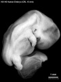
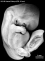
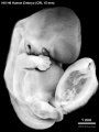
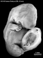
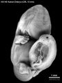
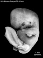
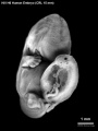
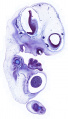
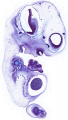
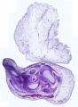
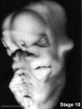
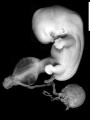
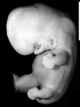
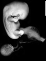
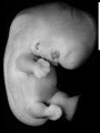
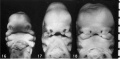
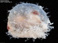
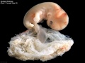
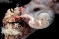
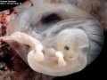
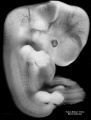
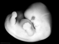
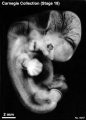
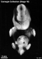
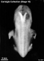
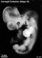
Pages and media in this category relate to Carnegie stage 18 (Week 7, 44 - 48 days, CRL 13 - 17 mm).
Subcategories
This category has the following 36 subcategories, out of 36 total.
C
- Carnegie Embryo 109
- Carnegie Embryo 144
- Carnegie Embryo 175
- Carnegie Embryo 1909
- Carnegie Embryo 2673
- Carnegie Embryo 296
- Carnegie Embryo 317
- Carnegie Embryo 351
- Carnegie Embryo 423
- Carnegie Embryo 424
- Carnegie Embryo 4430
- Carnegie Embryo 492
- Carnegie Embryo 511
- Carnegie Embryo 5747
- Carnegie Embryo 6522
- Carnegie Embryo 6524
- Carnegie Embryo 6525
- Carnegie Embryo 6528
- Carnegie Embryo 6529
- Carnegie Embryo 6533
- Carnegie Embryo 6551
- Carnegie Embryo 670
- Carnegie Embryo 719
- Carnegie Embryo 733
- Carnegie Embryo 7707
- Carnegie Embryo 8097
- Carnegie Embryo 8172
- Carnegie Embryo 8235
- Carnegie Embryo 8355
- Carnegie Embryo 841
- Carnegie Embryo 8812
- Carnegie Embryo 8945
- Carnegie Embryo 899
- Carnegie Embryo 9107
- Carnegie Embryo 9247
W
Pages in category 'Carnegie Stage 18'
The following 48 pages are in this category, out of 48 total.
C
- Template:Carnegie Collection stage 18 table
- Carnegie stage 18
- Template:Carnegie stage 18 links
- Template:CE109
- Template:CE144
- Template:CE1909
- Template:CE296
- Template:CE317
- Template:CE423
- Template:CE424
- Template:CE4430
- Template:CE492
- Template:CE511
- Template:CE535
- Template:CE6522
- Template:CE6524
- Template:CE6525
- Template:CE6527
- Template:CE6528
- Template:CE719
- Template:CE7707
- Template:CE8097
- Template:CE8172
- Template:CE8235
- Template:CE8355
- Template:CE841
- Template:CE9247
- Template:CS18
P
- Paper - Developmental horizons in human embryos stages 15-18
- Paper - Growth allometry of the myocardium in human embryos from stages 15 to 23
- Paper - Structural organization of the human cerebral cortex prior to the appearance of the cortical plate (1983)
- Paper - Studies on the development of the human larynx (1911)
- Paper - The human brain at stages 18-20 including the choroid plexuses and the amygdaloid and septal nuclei (1990)
R
Media in category 'Carnegie Stage 18'
The following 76 files are in this category, out of 76 total.
 Bailey336.jpg 912 × 597; 145 KB
 Carnegie stage 18 OPT.jpg 800 × 801; 43 KB
 Faulconer1951 fig03.jpg 1,014 × 847; 206 KB
 Faulconer1951 fig04.jpg 1,014 × 847; 215 KB
 HillH6 Stage 18 bf01.jpg 1,547 × 2,062; 274 KB
 HillH6 Stage 18 bf02.jpg 1,547 × 2,062; 337 KB
 HillH6 Stage 18 bf03.jpg 1,547 × 2,062; 287 KB
 HillH6 Stage 18 bf04.jpg 1,547 × 2,062; 318 KB
 HillH6 Stage 18 bf05.jpg 1,547 × 2,062; 218 KB
 HillH6 Stage 18 bf06.jpg 1,547 × 2,062; 232 KB
 HillH6 Stage 18 bf51.jpg 1,547 × 2,062; 198 KB
 Hinrichsen S226 001.jpg 1,280 × 2,246; 824 KB
 Hinrichsen S226 002.jpg 853 × 1,500; 394 KB
 Hinrichsen S226 003.jpg 1,102 × 1,500; 586 KB
 Hinrichsen S226 004.jpg 735 × 1,000; 269 KB
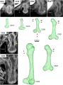 Human embryo femur CS18 to CS23.png 1,200 × 1,624; 1.42 MB
 Human embryo head stage18.jpg 300 × 400; 17 KB
 Human embryo head week 6 to 8.jpg 540 × 780; 66 KB
 Human Embryo Stage18-19.jpg 600 × 392; 17 KB
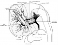
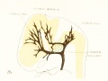
 Human embryonic renal branching 1.jpg 1,280 × 779; 236 KB
 Human embryonic tongue 08.jpg 650 × 470; 185 KB
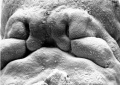
 Human stage18 face 01.jpg 500 × 504; 23 KB
 Human stage18 heart MRI - ventricular septation.jpg 1,014 × 652; 48 KB
 Keibel Mall 2 488.jpg 1,200 × 576; 317 KB
 Kyoto25783 stage18-dorsal.jpg 1,536 × 2,048; 152 KB
 Kyoto25783 stage18-left+yolk.jpg 1,536 × 2,048; 207 KB
 Kyoto25783 stage18-left.jpg 1,536 × 2,048; 226 KB
 Kyoto25783 stage18-right+yolk.jpg 1,536 × 2,048; 219 KB
 Kyoto25783 stage18-right.jpg 1,536 × 2,048; 228 KB
 Kyoto25783 stage18-ventral.jpg 1,536 × 2,048; 169 KB
 Lisser1911 fig01.jpg 1,119 × 900; 92 KB
 Lisser1911 fig02.jpg 1,119 × 900; 94 KB
 Lisser1911 fig03.jpg 1,119 × 900; 130 KB
 Lisser1911 fig04.jpg 1,119 × 900; 150 KB
 Lisser1911 fig05.jpg 1,119 × 900; 212 KB
 Lisser1911 fig06.jpg 1,119 × 900; 194 KB
 Lisser1911 fig07.jpg 1,477 × 886; 208 KB
 Lisser1911 fig08.jpg 1,477 × 886; 174 KB
 Lisser1911 fig09.jpg 1,477 × 886; 203 KB
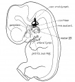
 Mall1906-fig21.jpg 1,000 × 763; 72 KB
 McClure1925 fig07.jpg 1,000 × 1,509; 185 KB
 McClure1925 fig08.jpg 900 × 1,245; 193 KB
 McClure1925 fig09.jpg 1,000 × 1,474; 193 KB
 McClure1925 fig10.jpg 1,000 × 1,437; 311 KB
 McClure1925 fig11.jpg 900 × 1,047; 185 KB
 McClure1925 fig12.jpg 1,000 × 1,564; 184 KB
 McClure1925 fig13.jpg 1,000 × 1,368; 331 KB
 Placental membranes.jpg 600 × 450; 99 KB
 Sensenig1951 plate02.jpg 2,078 × 2,619; 1.52 MB
 Sgalitzer1941 fig08.jpg 749 × 700; 60 KB
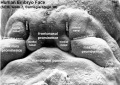
 Stage16-18 face.jpg 800 × 393; 34 KB
 Stage17-18 Primary palate.gif 458 × 283; 373 KB
 Stage18 bf1.jpg 1,000 × 750; 21 KB
 Stage18 bf10.jpg 1,200 × 897; 151 KB
 Stage18 bf11.jpg 1,200 × 900; 119 KB
 Stage18 bf12.jpg 1,200 × 800; 136 KB
 Stage18 bf13.jpg 1,200 × 900; 111 KB
 Stage18 bf14.jpg 1,518 × 2,000; 311 KB
 Stage18 bf1a.jpg 800 × 600; 15 KB
 Stage18 bf1b.jpg 600 × 450; 10 KB
 Stage18 bf1c.jpg 400 × 300; 5 KB
 Stage18 bf2.jpg 388 × 543; 28 KB
 Stage18 bf3.jpg 388 × 543; 23 KB
 Stage18 bf4.jpg 388 × 543; 20 KB
 Stage18 bf5.jpg 388 × 543; 24 KB
 Stage18 bf6.jpg 388 × 543; 26 KB
 Stage18 bf7.jpg 388 × 543; 22 KB
 Stage18 bf8.jpg 388 × 543; 22 KB
 Stage18 bf9.jpg 388 × 543; 24 KB
 Stage18 em01.jpg 800 × 569; 81 KB
 Stage18 em11.jpg 800 × 569; 99 KB
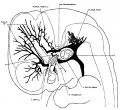
 Streeter1906 fig01.jpg 1,193 × 1,318; 167 KB
 Streeter1915 fig03.jpg 1,000 × 917; 139 KB
 Streeter1921 fig02.jpg 1,286 × 1,000; 138 KB
 Streeter1921 fig15.jpg 800 × 600; 54 KB
 Streeter1957 fig01.jpg 1,292 × 1,500; 218 KB


