Anatomy of the Human Body by Henry Gray: Difference between revisions
mNo edit summary |
mNo edit summary |
||
| Line 34: | Line 34: | ||
|} | |} | ||
{{Grays Embryo iBook table}} | {{Grays Embryo iBook table}} | ||
Revision as of 12:24, 22 April 2013
Introduction


Classic anatomy textbook widely reproduced online, particularly the anatomical illustrations, due to the fact that the 1918 edition is out of copyright. W.H. Lewis edited the 20th edition published in September 1918, the current 40th edition was published in 2008. The majority of images were anatomical drawings with some cartoon simplifications. The text also includes earlier historic drawings, particularly in the embryology section that commences the text.
Reference: Gray, Henry. Anatomy of the Human Body. Philadelphia: Lea & Febiger, 1918.
Clicking the Category:Gray's 1918 Anatomy should display a list of the images available on this current website. Note that over time the image naming has varied and requires better standardisation. Images used here may be altered and edited from those appearing in the original textbook.
| iBooks |
Anatomy of the Human Body on the Web for iPhone/iPad
| As an additional online educational project, I have also prepared 3 complete sets of images formatted specifically for the iPhone, and can be also used on the iPad, in different organisational layouts. Note the linked content below will look different when opened on other devices.
|

|
| iBook - Gray's Embryology | |
|---|---|

|
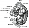
|
Images
Not all site images are included below. There may be several image versions (sizes, labeling, and formats gif, jpg, png).
| Historic Disclaimer - information about historic embryology pages |
|---|
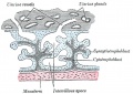
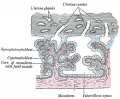
 |
1 Development
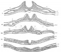
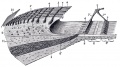
15 Neural Groove- series of sections dog embryo
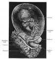
20 Dorsal human embryo 2.11 mm
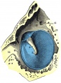
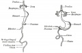
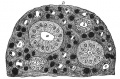
24 Diagram showing earliest observed stage of human ovum
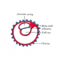
25 Diagram illustrating early formation of allantois and differentiation of body-stalk
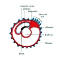
26 Diagram showing later stage of allantoic development with commencing constriction of the yolk-sac
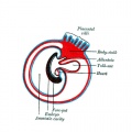
27 Diagram showing the expansion of amnion and delimitation of the umbilicus
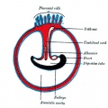
28 Diagram illustrating a later stage in the development of the umbilical cord
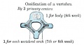
29
30

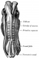
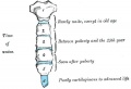
31 Model of human embryo 1.3 mm
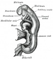
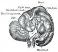
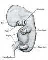
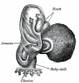
32 Human Embryo Day 8 to 9 (week 3)
33
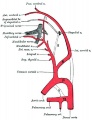
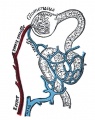
34 Uterus in the third and fourth month
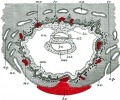
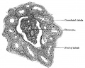
36 Primary Chorionic Villi
37 Secondary Chorionic Villi
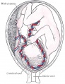
38 Fetus in Utero Between fifth and sixth months
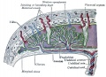
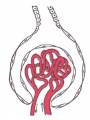
39 Placental circulation
40
52
53
58
60
63
65
83


101
118
119
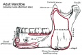
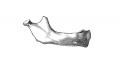
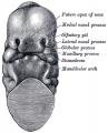
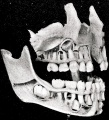
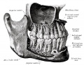
176 Human adult mandible
178 Human embryo CRL 24 mm outer aspect
179 Human embryo CRL 24 mm inner aspect
180 Human embryo CRL 95 mm outer aspect
181 Human embryo CRL 95 mm inner aspect
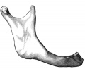
182 Mandible at birth
183 Mandible in childhood
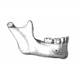
184 Mandible adult
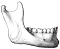
185 Mandible at old age


301-400
301
321
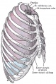
391 Adult human diaphragm (viewed from beneath)


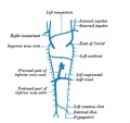
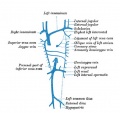
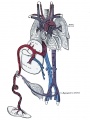
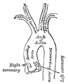
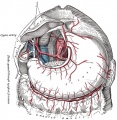
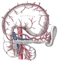
401-500 Cardiovascular
448 Artery and vein
467
468
470
472
474
475
476
477

478
479
480
492
498
502
506
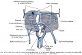
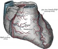
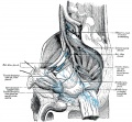
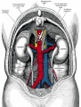
532 The celiac artery and its branches.
533 The celiac artery and its branches.
556


592 Lymphatic
592 Primary lymph sacs.
593 Lymph capillaries of the human conjunctiva.
594 Lymph capillaries from the human scrotum.
595 Lymph capillaries of the sole of the human foot.
596 Section through human tongue.
597 Lymph gland (Node).
598 Lymph gland tissue.
599 Thoracic and right lymphatic ducts.
600 Modes of origin of thoracic duct.
601 Terminal collecting trunks of right side.
602 Lymph glands of the head.
603 Lymphatics of pharynx.
604 Lymphatics of the face.
605 Lymphatics of the Tongue.
606 Lymph glands of the upper extremity.
607 Lymphatics of the mamma.
608 Lymphatic vessels of the dorsal hand surface.
609 Lymph glands of popliteal fossa.
610 Superficial lymph glands and vessels of the lower extremity.
611 Parietal lymph glands of the pelvis.
612 Iliopelvic glands.
613 Lymphatics of stomach.
614 Lymphatics of stomach.
615 Lymphatics of cecum and vermiform process.
616 Lymphatics of cecum and vermiform process.
617 Lymphatics of Colon.
618 Lymphatic of the Bladder.
619 Lymphatics of the Prostate.
620 Lymphatics of the Uterus.
621 Lymphatics of the thorax and abdomen.
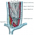
622 Tracheobronchial Lymph Glands


623 Neural
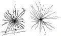
623 Neuroglia cells of brain.
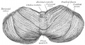
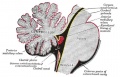
649 Human Fetal Hindbrain (3 months)
651 Human Embryo Brain (week 4.5 exterior view)
652 Human Embryo Brain (week 5 exterior view)
653 Human Embryo Brain (week 5 interior view)

654 Human Fetal Brain (3 months)
655 Human Fetal Brain (4 months)
658 Human Fetal Brain (5 months)
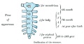
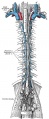
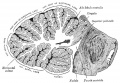
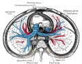
664 Transverse section of the medulla spinalis in the mid-thoracic region
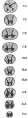
666 Transverse sections of the medulla spinalis at different levels
666 (new layout) Transverse sections of the medulla spinalis at different levels
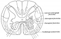
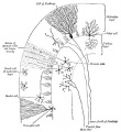
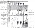
670 Diagram showing afferent (sensory) and efferent fibers
671 Spinal cord motor columns
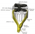
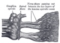
675 A spinal nerve with its anterior and posterior roots
677
678
697
698
702
704
705
706
708
715
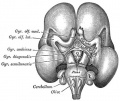
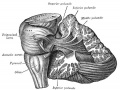
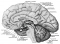
720 Median sagittal section of brain
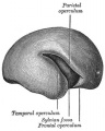
732
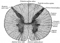
806


863 Vision
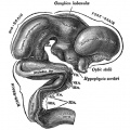
863 Chicken Optic Placode and Vesicle
864 Chicken Optic Placode and Vesicle
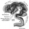
865 Human Optic Cup and Choroidal Fissure
866 Embryonic Rabbit Eye
867 Human Embryonic Eye
868 Section of developing eye of trout
869 Horizontal section of the eyeball
870 Cornea
871 Section of Human Cornea near the Margin
872 The Choroid and Iris
873 The Arteries of the Choroid and Iris
874 The Veins of the Choroid
875 Interior of anterior half of bulb of eye

876 Vessels of the choroid, ciliary processes, and iris of a child
877 Blood Vessels of the Eye
878 Iris front view
879 Interior of posterior half of bulb of left eye
880 Optic Nerve entering Eyeball
881 Section of Retina
882 Plan of Retinal Neurons
883 Section through front of Eyeball
884 Lens Structure
885 Lens
886 Lens Profile
887 Lens Epithelium
888 Sagittal section of right orbital cavity
889 Muscles of the right orbit
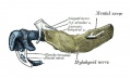
890 Right Ocular Muscles
891 Right Eye Fascia Bulbi
892 Front of left eye with eyelids separated to show medial canthus
893 Structure of the Eyelids
894 The tarsi and their ligaments
895 The Tarsal Glands
896 The Lacrimal Gland
897 Structures of the Lacrimal Gland


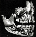
898 Hearing
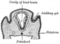
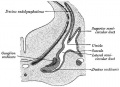
898 Section through human embryo head about twelve days old, in the region of the hind-brain
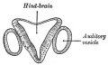
899 Section through hind-brain and auditory vesicles
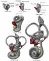
900 Lateral views of membranous labyrinth and acoustic complex
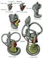
901 Median views of membranous labyrinth and acoustic complex in human embryos
902 Transverse section through head of fetal sheep in the region of the labyrinth
903 Transverse section of the cochlear duct of a fetal cat
904 Auricula or Pinna
905 Cranial surface of cartilage of right auricula
906 The muscles of the auricula
907 External and middle ear, opened from the front. Right side
908 Horizontal section through left ear; upper half of section.
909 Right tympanic membrane
910 Tympanic membrane
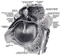
911 View of the inner wall of the tympanum
912 The right membrana tympani with the hammer and the chorda tympani, viewed from within, from behind, and from above.
913 Coronal section of right temporal bone
914 Right Tympanic Cavity Walls
915 Auditory Tube
916 Middle Ear - Malleus
917 Middle Ear - Incus
918 Middle Ear - Stapes
919 Chain of ossicles and their ligaments
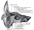
920 Right osseous labyrinth
921 Right osseous labyrinth, Interior.
922 Position of the right bony labyrinth in the skull
923 Inner Ear - Cochlea and Vestibule
924 Inner Ear - The Membranous Labyrinth
925 Right Human Membranous Labyrinth
926 Right Human Membranous Labyrinth
927 Human Semicircular Canal and Duct
928 Cross-section of Cochlea
929 Floor of Ductus Cochlearis
930 Limbus laminæ spiralis and membrana basilaris
931 Section through the spiral organ of Corti
932 The lamina reticularis and subjacent structures
933 Cochlear Division of the Acoustic Nerve


934 Somatosensory
934 End-bulb of Krause
935 Pacinian corpuscle
936 Papilla of the hand
937 Nerve ending of Ruffini
938 Organ of Golgi
939 Muscle Spindle


940 Integumentary
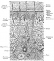
940 A diagrammatic sectional view of the skin
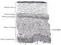
941 Section of Epidermis
942 The distribution of the bloodvessels in the skin of the sole of the foot
943 Longitudinal section through nail and its nail groove
944
945
946


947 Respiratory
947 The head and neck human embryo thirty-two days seen from the ventral surface.
948 Lung buds from a human embryo of about four weeks, showing commencing lobulations.
949 Lungs of a human embryo more advanced in development than week 4.
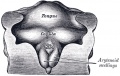
950 Cartilages of the larynx
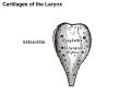
950 epiglottis cartilage
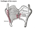
950 thyroid cartilage
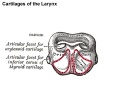
950 cricoid cartilage
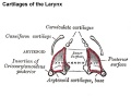
950 arytenoid cartilage
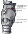
951 Ligaments of the larynx (anterior view)
952 Ligaments of the larynx (posterior view)
953 Larynx and upper part of the trachea
954
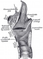
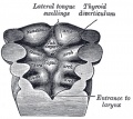
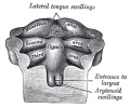
955 Larynx entrance
956
957
958
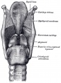
959
960
961 Cartilages of larynx, trachea, and bronchi (front view)
962 Bronchi and bronchioles
963
964
965
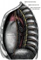
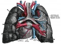
966 Lateral view of thorax, showing the relations of the pleuræ and lungs to the chest wall. Pleura in blue; lungs in purple.
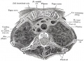
967 Transverse section through the upper margin of the second thoracic vertebra.
968
969
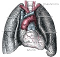
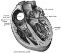
970 Front view of heart and lungs
971 Adult lungs
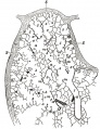
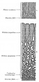
974 Lung secondary lobule
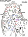
974 relabeled version
975 Lung primary lobule
975 relabeled version
976 Pig embryo lung


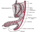
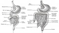
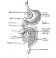
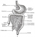
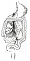
977 Gastrointestinal
977
978
979
980
981

982a

982b
983
984
985

986
987
987a
987b
988
989
990
991
992
993
994
996
997
998
999
1000

1001
1002
1003
1004
1005
1006
1007
1008
- Gray1009.jpg
1009
- Gray1010.jpg
1010

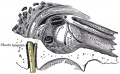
1027 Section through one of the crypts of the tonsil
1029 Front of nasa part of pharynx
1039
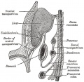
1095 Gall bladder and bile ducts laid open
1096 Gall bladder transverse section


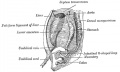
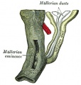
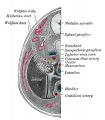
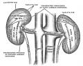
1108 Urogenital
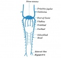
1108 Broad ligament of adult showing Epoöphoron
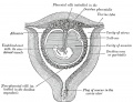
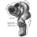
1109 Urogenital Sinus of Female Human Embryo of 8.5 to 9 weeks old
1111 Transverse section of Human Embryo 8.5 to 9 Weeks Old
1112 Longitudinal Section of Ovary of Cat Embryo of 9.4 cm long
1113 Section of the Ovary of a Newly Born Child
1114 Human Embryo (3.5 cm long) Testis Section of a Genital Cord
1115 Tail end of Human Embryo 25 to 29 Days Old
1116 Tail end of Human Embryo 32 to 33 Days Old
1117 Tail end of human embryo eight and a half to nine weeks old
1118 Primitive Kidney and Bladder
1119 Stages in the development of the external sexual organs in the male and female
1120 Abdomen
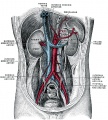
1121 Posterior abdominal wall

1122
1123
1124
1125
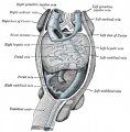
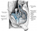
1126 Retroperitoneal structures

1126 Retroperitoneal structures
1127
1128
1129
1130
1131
1132
1133
1137
1138
1139
1140
1141
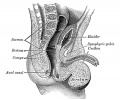
1152 Fundus of the bladder with the vesiculæ seminales
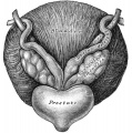
1153 Prostate and seminal vesicles
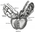
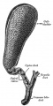
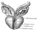
1160 Prostate Gland
- Gray1173.jpg
1173


1174 Endocrine
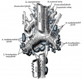
The Ductless Glands
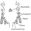
1174 The thyroid gland and its relations.
1175
1181 Pituitary - Median sagittal hypophysis adult monkey
1192


1193 Surface Anatomy
Surface Anatomy and Surface Markings
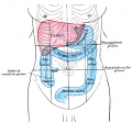
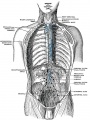
Abdomen Surface Markings for Liver, Stomach, and Great Intestine
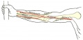
Front of right upper extremity, showing surface markings for bones and nerves.
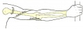
Back of right upper extremity, showing surface markings for bones and nerves.
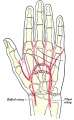
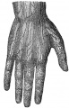
Left Hand Palm, position of skin creases and bones, and surface markings for the volar arches


Glossary Links
- Glossary: A | B | C | D | E | F | G | H | I | J | K | L | M | N | O | P | Q | R | S | T | U | V | W | X | Y | Z | Numbers | Symbols | Term Link
Cite this page: Hill, M.A. (2024, May 28) Embryology Anatomy of the Human Body by Henry Gray. Retrieved from https://embryology.med.unsw.edu.au/embryology/index.php/Anatomy_of_the_Human_Body_by_Henry_Gray
- © Dr Mark Hill 2024, UNSW Embryology ISBN: 978 0 7334 2609 4 - UNSW CRICOS Provider Code No. 00098G