Advanced - Cardiac Septation: Difference between revisions
No edit summary |
mNo edit summary |
||
| (6 intermediate revisions by the same user not shown) | |||
| Line 6: | Line 6: | ||
All of the partitioning of the primitive heart occurs between the middle of the fourth week and the end of the fifth week. Division of the atrioventricular canal is described below while septation of the atria and ventricles is described | All of the partitioning of the primitive heart occurs between the middle of the fourth week and the end of the fifth week. Division of the atrioventricular canal is described below while [[Advanced_-_Cardiac_Septation_2|'''septation of the atria and ventricles''' is described here]]. | ||
===Division of the AV Canal=== | ===Division of the AV Canal=== | ||
Two endocardial cushions form on the dorsal and ventral surfaces of the AV canal. Following expansion of the cardiac jelly, epithelial to mesenchymal transformation (EMT) of the endocardial cells in the canal occurs forming the cushions. Synergistic signalling between BMP and TGFβ facilitates EMT. The cushions grow as they are invaded by mesenchymal cells from the endocardium during the fifth week, eventually fusing to create the right and left AV canals, hence partially separating the primitive atrium and ventricle | Two endocardial cushions form on the dorsal and ventral surfaces of the AV canal. Following expansion of the cardiac jelly, epithelial to mesenchymal transformation (EMT) of the endocardial cells in the canal occurs forming the cushions. Synergistic signalling between BMP and TGFβ facilitates EMT. The cushions grow as they are invaded by mesenchymal cells from the endocardium during the fifth week, eventually fusing to create the right and left AV canals, hence partially separating the primitive atrium and ventricle (Click image to play on current page or [[Media:Heart septation 001.mp4|Play video on new page]]). | ||
<html5media height="720" width="560">File:Heart septation 001.mp4</html5media> | |||
{| width="100%" | {| width="100%" | ||
Latest revision as of 00:36, 18 June 2014
| Begin Advanced | Heart Fields | Heart Tubes | Cardiac Looping | Cardiac Septation | Outflow Tract | Valve Development | Cardiac Conduction | Cardiac Abnormalities | Molecular Development |
| Cardiac Embryology | Begin Basic | Begin Intermediate | Begin Advanced |
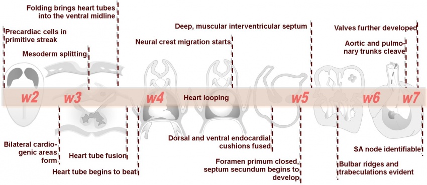
All of the partitioning of the primitive heart occurs between the middle of the fourth week and the end of the fifth week. Division of the atrioventricular canal is described below while septation of the atria and ventricles is described here.
Division of the AV Canal
Two endocardial cushions form on the dorsal and ventral surfaces of the AV canal. Following expansion of the cardiac jelly, epithelial to mesenchymal transformation (EMT) of the endocardial cells in the canal occurs forming the cushions. Synergistic signalling between BMP and TGFβ facilitates EMT. The cushions grow as they are invaded by mesenchymal cells from the endocardium during the fifth week, eventually fusing to create the right and left AV canals, hence partially separating the primitive atrium and ventricle (Click image to play on current page or Play video on new page).
<html5media height="720" width="560">File:Heart septation 001.mp4</html5media>
| Back to Looping | Next: Cardiac Septation 2 | |
| Go to this section in the intermediate level |
