ACPS Seminar 2014 - Implantation: Difference between revisions
mNo edit summary |
|||
| Line 51: | Line 51: | ||
HLA-G expression (red) on trophoblast cell columns (CC) and extravillous trophoblasts and not seen for syncitiotrophoblast (SYN) cells.<ref><pubmed>22438923</pubmed>| [http://www.plosone.org/article/info%3Adoi%2F10.1371%2Fjournal.pone.0033395 PLoS One.]</ref> | HLA-G expression (red) on trophoblast cell columns (CC) and extravillous trophoblasts and not seen for syncitiotrophoblast (SYN) cells.<ref><pubmed>22438923</pubmed>| [http://www.plosone.org/article/info%3Adoi%2F10.1371%2Fjournal.pone.0033395 PLoS One.]</ref> | ||
==Protein Expression | ==Analysis== | ||
{| | |||
| [[File:Sandwich ELISA.jpg|400px]] | |||
| [[File:Microarray_technology.jpg|400px]] | |||
|- | |||
| Protein Expression | |||
| mRNA Expression | |||
|} | |||
==External Links== | ==External Links== | ||
Revision as of 14:00, 17 March 2014
Understanding Implantation and the new Uterine Tube Biobank
|

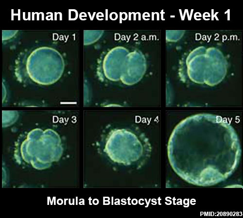
Human Development - Week 1

|
<mediaplayer width='500' height='450' image="http://embryology.med.unsw.edu.au/embryology/images/0/07/Human_blastocyst_day_1-5.jpg">File:Human_blastocyst_day_3-6.mp4</mediaplayer>
|
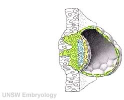
Human Development - Week 2
| <mediaplayer width='250' height='260' image="http://php.med.unsw.edu.au/embryology/images/a/a9/Week2_001_icon.jpg">File:Week2_001.mp4</mediaplayer> | This animation shows the process of implantation, occurring during week 2 (GA week 4) of development in humans. The beginning of the animation shows adplantation to the the uterus lining (endometrium epithelium). The hatched blastocyst with a flat outer layer of trophoblast cells (green), the inner cell mass which has formed into the bilaminar embryo (epiblast and hypoblast) and the large fluid-filled space (blastocoel).
|
Tube Biobank

HLA-G

HLA-G expression (red) on trophoblast cell columns (CC) and extravillous trophoblasts and not seen for syncitiotrophoblast (SYN) cells.[2]
Analysis

|

|
| Protein Expression | mRNA Expression |
External Links
External Links Notice - The dynamic nature of the internet may mean that some of these listed links may no longer function. If the link no longer works search the web with the link text or name. Links to any external commercial sites are provided for information purposes only and should never be considered an endorsement. UNSW Embryology is provided as an educational resource with no clinical information or commercial affiliation.
References
- ↑ <pubmed>20890283</pubmed>| Nat Biotechnol.
- ↑ <pubmed>22438923</pubmed>| PLoS One.
Cite this page: Hill, M.A. (2024, May 17) Embryology ACPS Seminar 2014 - Implantation. Retrieved from https://embryology.med.unsw.edu.au/embryology/index.php/ACPS_Seminar_2014_-_Implantation
- © Dr Mark Hill 2024, UNSW Embryology ISBN: 978 0 7334 2609 4 - UNSW CRICOS Provider Code No. 00098G