2010 Lecture 5: Difference between revisions
No edit summary |
|||
| Line 18: | Line 18: | ||
'''Coelom''', meaning "cavity", and major fluid-filled cavities can be seen to form both within the embryo (intraembryonic coelom) and outside the embryo (extraembryonic coelom). The '''intraembryonic coelom''' is the single primitive cavity that lies within the mesoderm layer that will eventually form the 3 major anatomical body cavities ('''pericardial, pleural, peritoneal'''). | '''Coelom''', meaning "cavity", and major fluid-filled cavities can be seen to form both within the embryo (intraembryonic coelom) and outside the embryo (extraembryonic coelom). The '''intraembryonic coelom''' is the single primitive cavity that lies within the mesoderm layer that will eventually form the 3 major anatomical body cavities ('''pericardial, pleural, peritoneal'''). | ||
==UNSW Embryology Links== | |||
* '''Mesoderm Slides''' [[2009_Lecture_5|2009 Lecture]] | [http://embryology.med.unsw.edu.au/pdf/ANAT2341L6Mesoderms1.pdf Mesoderm Lecture 2008 - 1 slide/page ] | [http://embryology.med.unsw.edu.au/pdf/ANAT2341L6Mesoderms4.pdf Lecture 3 2008 Slides - 4 slides/page] | [http://embryology.med.unsw.edu.au/pdf/ANAT2341L6Mesoderms6.pdf Mesoderm Lecture 2008 Slides - 6 slides/page] | |||
* '''Mesoderm''' [http://embryology.med.unsw.edu.au/Movies/week4.htm Week 4 Movies] | [http://embryology.med.unsw.edu.au/Movies/mesoderm.htm Mesoderm Movies] | | |||
* '''Mesoderm Notes''' [http://embryology.med.unsw.edu.au/Notes/week4.htm Week 4] | [http://embryology.med.unsw.edu.au/Notes/week4_4.htm Week 4 - Somites] | [http://embryology.med.unsw.edu.au/Notes/coelom.htm Coelomic Cavity Development] | [http://embryology.med.unsw.edu.au/Notes/skmus.htm Musculoskeletal Development] | | |||
{{Template:2010ANAT2341}} | {{Template:2010ANAT2341}} | ||
Revision as of 09:03, 7 August 2010
Mesoderm Development
Introduction
| File:Mesoderm_001_icon.jpg</wikiflv> | This animation shows the migration of mesoderm throughout the embryonic disc during gastrulation. |
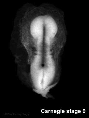
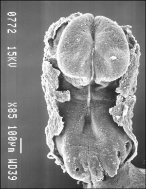
We have seen the following processes during early human development so far: fertilization and blastocyst development in the first week, implantation in the second week, early placentation and bilaminar to trilaminar in the third week. In the third to fourth week we will now follow the development of the trilaminar embryo as each layer begins to differentiate into the primordia of different tissues within the embryo. From this point onward the lectures will not be in a strict timeline format as we will have to follow each layer (ectoderm, mesoderm, endoderm) forward through its early development, and then jump back to discuss the next layer.
This lecture will look at mesoderm development and formation of the body cavities.
Mesoderm means the "middle layer" and it is from this layer that nearly all the bodies connective tissues are derived. In early mesoderm development a number of transient structures will form and then be lost as tissue structure is patterned and organised. Humans are vertebrates, with a "backbone", and the first mesoderm structure we will see form after the notochord will be somites.
Coelom, meaning "cavity", and major fluid-filled cavities can be seen to form both within the embryo (intraembryonic coelom) and outside the embryo (extraembryonic coelom). The intraembryonic coelom is the single primitive cavity that lies within the mesoderm layer that will eventually form the 3 major anatomical body cavities (pericardial, pleural, peritoneal).
UNSW Embryology Links
- Mesoderm Slides 2009 Lecture | Mesoderm Lecture 2008 - 1 slide/page | Lecture 3 2008 Slides - 4 slides/page | Mesoderm Lecture 2008 Slides - 6 slides/page
- Mesoderm Week 4 Movies | Mesoderm Movies |
- Mesoderm Notes Week 4 | Week 4 - Somites | Coelomic Cavity Development | Musculoskeletal Development |
Glossary Links
- Glossary: A | B | C | D | E | F | G | H | I | J | K | L | M | N | O | P | Q | R | S | T | U | V | W | X | Y | Z | Numbers | Symbols | Term Link
Course Content 2010
Embryology Introduction | Cell Division/Fertilization | Lab 1 | Week 1&2 Development | Week 3 Development | Lab 2 | Mesoderm Development | Ectoderm, Early Neural, Neural Crest | Lab 3 | Early Vascular Development | Placenta | Lab 4 | Endoderm, Early Gastrointestinal | Respiratory Development | Lab 5 | Head Development | Neural Crest Development | Lab 6 | Musculoskeletal Development | Limb Development | Lab 7 | Kidney | Genital | Lab 8 | Sensory | Stem Cells | Stem Cells | Endocrine | Lab 10 | Late Vascular Development | Integumentary | Lab 11 | Birth, Postnatal | Revision | Lab 12 | Lecture Audio | Course Timetable
Cite this page: Hill, M.A. (2024, April 27) Embryology 2010 Lecture 5. Retrieved from https://embryology.med.unsw.edu.au/embryology/index.php/2010_Lecture_5
- © Dr Mark Hill 2024, UNSW Embryology ISBN: 978 0 7334 2609 4 - UNSW CRICOS Provider Code No. 00098G

