2010 Lecture 11: Difference between revisions
No edit summary |
No edit summary |
||
| Line 21: | Line 21: | ||
* '''Larsen’s Human Embryology''' by GC. Schoenwolf, SB. Bleyl, PR. Brauer and PH. Francis-West - Chapter 12 Development of the Head, the Neck, the Eyes, and the Ears pp349 - 418. | * '''Larsen’s Human Embryology''' by GC. Schoenwolf, SB. Bleyl, PR. Brauer and PH. Francis-West - Chapter 12 Development of the Head, the Neck, the Eyes, and the Ears pp349 - 418. | ||
{| | |||
| <Flowplayer width="420" height="500" autoplay="true">Face_001.flv</Flowplayer> | |||
| | |||
'''Development of the Face''' | |||
This animation shows a ventral view of development of the human face from approximately week 5 through to neonate. | |||
The separate embryonic components that contribute to the face have been colour coded. | |||
* '''Frontonasal Prominence (white)''' | |||
* <font color=blueviolet>'''Frontonasal Prominence - Lateral nasal'''</font> (purple) | |||
* <font color=yellowgreen>'''Frontonasal Prominence - Medial nasal'''</font> (green) | |||
* <font color=gold>'''Pharyngeal Arch 1 - Maxillary prominence'''</font> (yellow) | |||
* <font color=darkorange>'''Pharyngeal Arch 1 - Mandibular prominence'''</font> (orange) | |||
* '''Stomodeum (black)''' | |||
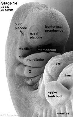
The [[S#stomodeum|stomodeum]] is the primordial mouth region and a surface central depression lying between the forebrain bulge and the heart bulge. At the floor of the stomodeum indentation is the [[B#buccopharyngeal membrane|buccopharyngeal membrane]] (oral membrane). | |||
Note the complex origin of the maxillary region (upper jaw) requiring the fusion of several embryonic elements, abnormalities of this process lead to [[C#cleft lip|cleft lip]] and [[C#cleft palate|cleft palate]]. | |||
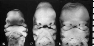
See also the movie ([[Quicktime_Movie_-_Stage_16_to_18_Face|Quicktime]] | [[Quicktime_Movie_-_Stage_16_to_18_Face|Flash]]) showing a similar view of human embryo faces between [[Carnegie stage 16|Carnegie stage 16]] to [[Carnegie stage 18|18]]. | |||
'''Links:''' [[Quicktime Development Animation - Face]] | [[Media:Face_001.mov|Quicktime version]] | [[Head Development]] | |||
|} | |||
{{Template:2010ANAT2341}} | {{Template:2010ANAT2341}} | ||
[[Category:2010ANAT2341]][[Category:Science-Undergraduate]] [[Category:Head]] ] [[Category:Pharyngeal Arch]] | [[Category:2010ANAT2341]][[Category:Science-Undergraduate]] [[Category:Head]] ] [[Category:Pharyngeal Arch]] | ||
Revision as of 09:26, 27 August 2010
Head Development
Introduction
The face is the anatomical feature which is truly unique to each human, though the basis of its general development is identical for all humans and similar to that seem for other species. The face has a complex origin arising from a number of head structures and sensitive to a number of teratogens during critical periods of its development. The related structures of upper lip and palate significantly contribute to the majority of face abnormalities.
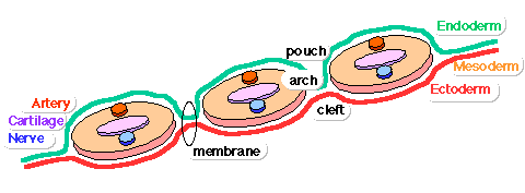
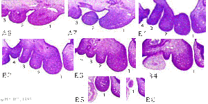
The head and neck structures are more than just the face, and are derived from pharyngeal arches 1 - 6 with the face forming from arch 1 and 2 and the frontonasal prominence. Each arch contains similar Arch components derived from endoderm, mesoderm, neural crest and ectoderm. These components though will form different structures depending on their arch origin. Because the head contains many different structures also review notes on Special Senses (Sensory System Development), Respiratation ([[Respiratory System Development]), Integumentary (Teeth), Endocrine (Endocrine System Development thyroid, pituitary) and Ultrasound - Cleft lip/palate.
Lecture Objectives
- List the main structures derived from the pharyngeal arches, pouches and clefts.
- Know the stages and structures involved in the development of the face.
- Predict the results of abnormal development of the face and palate.
- Briefly summarise the development of the tongue.
Textbook References
- The Developing Human: Clinically Oriented Embryology (8th Edition) by Keith L. Moore and T.V.N Persaud - Moore & Persaud Chapter Chapter 10 The Pharyngeal Apparatus pp201 - 240.
- Larsen’s Human Embryology by GC. Schoenwolf, SB. Bleyl, PR. Brauer and PH. Francis-West - Chapter 12 Development of the Head, the Neck, the Eyes, and the Ears pp349 - 418.
| <Flowplayer width="420" height="500" autoplay="true">Face_001.flv</Flowplayer> |
Development of the Face
The separate embryonic components that contribute to the face have been colour coded.
The stomodeum is the primordial mouth region and a surface central depression lying between the forebrain bulge and the heart bulge. At the floor of the stomodeum indentation is the buccopharyngeal membrane (oral membrane). Note the complex origin of the maxillary region (upper jaw) requiring the fusion of several embryonic elements, abnormalities of this process lead to cleft lip and cleft palate. See also the movie (Quicktime | Flash) showing a similar view of human embryo faces between Carnegie stage 16 to 18.
|
Glossary Links
- Glossary: A | B | C | D | E | F | G | H | I | J | K | L | M | N | O | P | Q | R | S | T | U | V | W | X | Y | Z | Numbers | Symbols | Term Link
Course Content 2010
Embryology Introduction | Cell Division/Fertilization | Lab 1 | Week 1&2 Development | Week 3 Development | Lab 2 | Mesoderm Development | Ectoderm, Early Neural, Neural Crest | Lab 3 | Early Vascular Development | Placenta | Lab 4 | Endoderm, Early Gastrointestinal | Respiratory Development | Lab 5 | Head Development | Neural Crest Development | Lab 6 | Musculoskeletal Development | Limb Development | Lab 7 | Kidney | Genital | Lab 8 | Sensory | Stem Cells | Stem Cells | Endocrine | Lab 10 | Late Vascular Development | Integumentary | Lab 11 | Birth, Postnatal | Revision | Lab 12 | Lecture Audio | Course Timetable
Cite this page: Hill, M.A. (2024, May 2) Embryology 2010 Lecture 11. Retrieved from https://embryology.med.unsw.edu.au/embryology/index.php/2010_Lecture_11
- © Dr Mark Hill 2024, UNSW Embryology ISBN: 978 0 7334 2609 4 - UNSW CRICOS Provider Code No. 00098G ]


