2010 BGD Lecture - Development of the Embryo/Fetus 2: Difference between revisions
| Line 11: | Line 11: | ||
* Dynamic changes internal and external | * Dynamic changes internal and external | ||
* Carnegie stages illustrate external development | * Carnegie stages illustrate external development | ||
==First 8 Weeks== | |||
[[File:Human_Carnegie_stage_1-23.jpg]] | |||
==Week 3== | |||
==Somitogenesis== | |||
[[File:Stage_9_SEM1.jpg|thumb|stage 9 Embryo]] [[File:Stage10 bf6b.jpg|thumb|stage 10 Embryo]] | |||
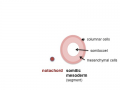
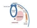
'''Mesoderm''' means the "middle layer" and it is from this layer that nearly all the bodies connective tissues are derived. In early mesoderm development a number of transient structures will form and then be lost as tissue structure is patterned and organised. Humans are vertebrates, with a "backbone", and the first mesoderm structure we will see form after the notochord will be '''[[S#somite|somites]]'''. | |||
'''Coelom''', meaning "cavity", and major fluid-filled cavities can be seen to form both within the embryo ([[I#intraembryonic coelom|intraembryonic coelom]]) and outside the embryo (extraembryonic coelom). The '''intraembryonic coelom''' is the single primitive cavity that lies within the mesoderm layer that will eventually form the 3 major anatomical body cavities ('''[[P#pericardial cavity|pericardial]], [[P#pleural cavity|pleural]], [[P#peritoneal cavity|peritoneal]]'''). | |||
<gallery> | |||
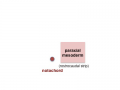
Image:Mesoderm cartoon1.gif|trilaminar embryo | |||
Image:Mesoderm cartoon2.gif|mesoderm regions | |||
Image:Mesoderm cartoon3.gif|somite coelom | |||
Image:Mesoderm cartoon4.gif|neural tube and neural crest | |||
</gallery> | |||
[[File:Stage11 sem100c.jpg||thumb|stage 11 Embryo]] | |||
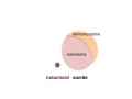
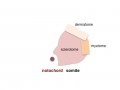
Somite initially forms 2 main components | |||
* ventromedial- sclerotome forms vertebral body and intervertebral disc | |||
* dorsolateral - dermomyotome forms dermis and skeletal muscle | |||
<gallery> | |||
Image:Somite cartoon1.png|paraxial mesoderm | |||
Image:Somite cartoon2.png|early somite | |||
Image:Somite cartoon3.png|sclerotome and dermomyotome | |||
Image:Somite cartoon4.png|dermatome and myotome | |||
Image:Somite cartoon5.png|epaxial and hypaxial muscles | |||
</gallery> | |||
[[File:Somite 001 icon.jpg|90px|link=Development_Animation_-_Somite_Musculoskeletal]] | |||
==Heart== | ==Heart== | ||
Revision as of 21:11, 10 May 2010
Introduction
--Mark Hill 20:54, 3 May 2010 (EST) I am currently updating the notes that will appear here for 2010. Previous 2008 Lecture
This lecture covers the period of Embryonic development, in Humans from week 3 to week 8 and is divided into 23 Carnegie stages of embryonic development. There will also be a brief introduction to fetal development.
This period of development will also be covered in your Practical, which is currently being updated for 2010 and will be available online. Note, the period from week 9 to week 38 is considered Fetal development and will be covered in detail in the Laboratory 11.
- Embryonic Development
- Organ and System formation (Functioning / Not Functional)
- Dynamic changes internal and external
- Carnegie stages illustrate external development
First 8 Weeks

Week 3
Somitogenesis


Mesoderm means the "middle layer" and it is from this layer that nearly all the bodies connective tissues are derived. In early mesoderm development a number of transient structures will form and then be lost as tissue structure is patterned and organised. Humans are vertebrates, with a "backbone", and the first mesoderm structure we will see form after the notochord will be somites.
Coelom, meaning "cavity", and major fluid-filled cavities can be seen to form both within the embryo (intraembryonic coelom) and outside the embryo (extraembryonic coelom). The intraembryonic coelom is the single primitive cavity that lies within the mesoderm layer that will eventually form the 3 major anatomical body cavities (pericardial, pleural, peritoneal).

trilaminar embryo
mesoderm regions
somite coelom

neural tube and neural crest


Somite initially forms 2 main components
- ventromedial- sclerotome forms vertebral body and intervertebral disc
- dorsolateral - dermomyotome forms dermis and skeletal muscle
paraxial mesoderm
early somite
sclerotome and dermomyotome
dermatome and myotome
epaxial and hypaxial muscles


![]()
Heart
.jpg)
Head

Upper and Lower Limb


Limb development occurs at different times for forelimbs and hindlimbs. In the mid-4th week, human upper limb buds first form and lower limbs about 2 days later. The limbs form at vertebra segmental levels C5-C8 (upper limbs) L3-L5 (lower limbs).
Glossary Links
- Glossary: A | B | C | D | E | F | G | H | I | J | K | L | M | N | O | P | Q | R | S | T | U | V | W | X | Y | Z | Numbers | Symbols | Term Link
- 2010 BGD: Lecture 1 | Lecture 2 | Practical 3 | Practical 6 | Practical 12
Cite this page: Hill, M.A. (2024, May 17) Embryology 2010 BGD Lecture - Development of the Embryo/Fetus 2. Retrieved from https://embryology.med.unsw.edu.au/embryology/index.php/2010_BGD_Lecture_-_Development_of_the_Embryo/Fetus_2
- © Dr Mark Hill 2024, UNSW Embryology ISBN: 978 0 7334 2609 4 - UNSW CRICOS Provider Code No. 00098G