2009 Lecture 17: Difference between revisions
No edit summary |
|||
| Line 1: | Line 1: | ||
=Sensory Development - Hearing= | =Sensory Development - Hearing= | ||
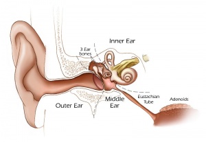
[[File:Hearing cartoon.jpg|300px|left]]We use the sense of balance and hearing to position ourselves in space, sense our surrounding environment, and to communicate. Portions of the ear appear very early in development as specialized region (otic placode) on the embryo surface that sinks into the mesenchyme to form a vesicle (otic vesicle = otocyst) that form the inner ear. | [[File:Hearing cartoon.jpg|300px|left]]We use the sense of balance and hearing to position ourselves in space, sense our surrounding environment, and to communicate. Portions of the ear appear very early in development as specialized region (otic placode) on the embryo surface that sinks into the mesenchyme to form a vesicle (otic vesicle = otocyst) that form the inner ear. | ||
This region connects centrally to the nervous system and peripherally through specialized bones to the external ear (auricle). This organisation develops different sources forming the 3 ear parts: inner ear (otic placode, otocyst), middle ear (1st pharyngeal pouch and 1st and 2nd arch mesenchyme), and outer ear (1st pharyngeal cleft and 6 surface hillocks). | |||
This complex origin, organisation, and timecourse means that abnormal development of any one system can impact upon the development of hearing. | |||
{{Template:2009ANAT2341}} | {{Template:2009ANAT2341}} | ||
[[Category:2009ANAT2341]][[Category:Science-Undergraduate]] | [[Category:2009ANAT2341]][[Category:Science-Undergraduate]] | ||
Revision as of 21:36, 24 September 2009
Sensory Development - Hearing
We use the sense of balance and hearing to position ourselves in space, sense our surrounding environment, and to communicate. Portions of the ear appear very early in development as specialized region (otic placode) on the embryo surface that sinks into the mesenchyme to form a vesicle (otic vesicle = otocyst) that form the inner ear.
This region connects centrally to the nervous system and peripherally through specialized bones to the external ear (auricle). This organisation develops different sources forming the 3 ear parts: inner ear (otic placode, otocyst), middle ear (1st pharyngeal pouch and 1st and 2nd arch mesenchyme), and outer ear (1st pharyngeal cleft and 6 surface hillocks).
This complex origin, organisation, and timecourse means that abnormal development of any one system can impact upon the development of hearing.
Glossary Links
- Glossary: A | B | C | D | E | F | G | H | I | J | K | L | M | N | O | P | Q | R | S | T | U | V | W | X | Y | Z | Numbers | Symbols | Term Link
Course Content 2009
Embryology Introduction | Cell Division/Fertilization | Cell Division/Fertilization | Week 1&2 Development | Week 3 Development | Lab 2 | Mesoderm Development | Ectoderm, Early Neural, Neural Crest | Lab 3 | Early Vascular Development | Placenta | Lab 4 | Endoderm, Early Gastrointestinal | Respiratory Development | Lab 5 | Head Development | Neural Crest Development | Lab 6 | Musculoskeletal Development | Limb Development | Lab 7 | Kidney | Genital | Lab 8 | Sensory - Ear | Integumentary | Lab 9 | Sensory - Eye | Endocrine | Lab 10 | Late Vascular Development | Fetal | Lab 11 | Birth, Postnatal | Revision | Lab 12 | Lecture Audio | Course Timetable
Cite this page: Hill, M.A. (2024, May 18) Embryology 2009 Lecture 17. Retrieved from https://embryology.med.unsw.edu.au/embryology/index.php/2009_Lecture_17
- © Dr Mark Hill 2024, UNSW Embryology ISBN: 978 0 7334 2609 4 - UNSW CRICOS Provider Code No. 00098G
