Category:Renal
From Embryology
This Embryology category shows media and pages related to renal system development. This includes the kidney, ureters, urinary bladder and urethra.
Subcategories
This category has the following 4 subcategories, out of 4 total.
Pages in category 'Renal'
The following 5 pages are in this category, out of 205 total.
(previous page) (next page)(previous page) (next page)
Media in category 'Renal'
The following 200 files are in this category, out of 283 total.
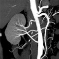
(previous page) (next page)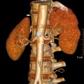 Accessory renal artery.jpg 800 × 798; 103 KB
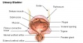 Adult bladder.jpg 520 × 273; 34 KB
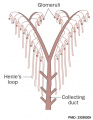 Adult nephron distribution.jpg 502 × 649; 56 KB
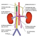 Adult renal venous cartoon.jpg 600 × 600; 62 KB
 Adult urinary bladder.jpg 1,164 × 1,000; 148 KB
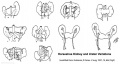
 Australian abnormalities 81-92 urogenital.jpg 600 × 429; 54 KB
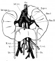
 Bailey305.jpg 931 × 440; 82 KB
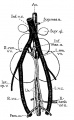
 Bailey306.jpg 624 × 419; 35 KB
 Bailey307.jpg 832 × 833; 128 KB
 Bailey308.jpg 791 × 848; 88 KB
 Bailey309.jpg 594 × 592; 58 KB
 Bailey311.jpg 758 × 578; 50 KB
 Bailey312.jpg 823 × 839; 82 KB
 Bailey313.jpg 543 × 649; 79 KB
 Bailey314.jpg 455 × 461; 25 KB
 Bailey315.jpg 689 × 672; 121 KB
 Bailey316.jpg 671 × 325; 58 KB
 Bailey317-319.jpg 663 × 1,080; 99 KB
 Bailey320.jpg 869 × 582; 75 KB
 Bailey321.jpg 596 × 671; 77 KB
 Bailey322.jpg 766 × 476; 62 KB
 Bailey323.jpg 653 × 521; 50 KB
 Bailey324 325.jpg 706 × 888; 114 KB
 Bailey324.jpg 913 × 558; 78 KB
 Bailey325.jpg 876 × 636; 101 KB
 Bailey326.jpg 502 × 512; 45 KB
 Bailey327.jpg 872 × 567; 89 KB
 Bailey333.jpg 830 × 445; 74 KB
 Bailey340.jpg 814 × 657; 62 KB
 Bladder exstrophy - female.jpg 1,200 × 900; 112 KB
 Bladder exstrophy - male.jpg 637 × 480; 31 KB
 Bladder Exstrophy.jpg 600 × 383; 37 KB
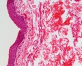
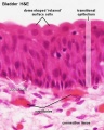
 Bladder histology 001.jpg 1,280 × 1,024; 522 KB
 Bladder histology 002.jpg 1,280 × 1,024; 295 KB
 Bladder histology 003.jpg 1,280 × 1,024; 229 KB
 Bladder histology 004.jpg 1,280 × 1,024; 212 KB
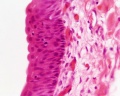
 Bladder histology 01.jpg 480 × 600; 29 KB
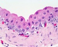
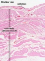 Bladder histology.jpg 300 × 400; 56 KB
 Boyden1931 fig02.jpg 773 × 860; 75 KB
 Boyden1931 fig03.jpg 541 × 878; 56 KB
 Boyden1931 fig04.jpg 1,280 × 652; 130 KB
 Boyden1931 fig05.jpg 1,000 × 646; 97 KB
 Caudal duplication syndrome.jpg 700 × 599; 47 KB
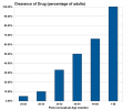 Drug-clearance-rates.png 652 × 542; 14 KB
 Eisendrath1925 fig01.jpg 1,000 × 882; 52 KB
 Eisendrath1925 fig02a.jpg 1,000 × 1,184; 105 KB
 Eisendrath1925 fig04.jpg 1,000 × 983; 85 KB
 Eisendrath1925 fig16.jpg 1,000 × 1,287; 76 KB
 Embryo renal venous cartoon.jpg 600 × 600; 68 KB
 Female genital and ureter abnormality 01.jpg 766 × 732; 86 KB
 Female genital and ureter abnormality 02.jpg 766 × 733; 78 KB
 Female genital and ureter abnormality 03.jpg 766 × 762; 79 KB
 Fetal 10wk urogenital 1.jpg 800 × 600; 109 KB
 Fetal 10wk urogenital 2.jpg 800 × 600; 110 KB
 Fetal 10wk urogenital 3.jpg 800 × 600; 107 KB
 Fetal 10wk urogenital 4.jpg 800 × 600; 105 KB
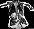
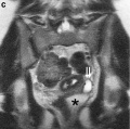
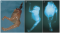
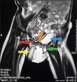
 Fetal kidney MRI 01.jpg 797 × 880; 68 KB
 Fetal kidney MRI 02.jpg 797 × 880; 64 KB
 Fetal kidney.jpg 689 × 623; 43 KB
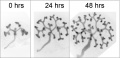
 Fetal nephron development 01.jpg 1,338 × 689; 119 KB
 Girgis09.jpg 342 × 1,033; 54 KB
 Glomerular podocyte cartoon 02.jpg 1,000 × 280; 51 KB
 Glomerular podocyte cartoon.jpg 800 × 199; 19 KB
 Gray0585.jpg 752 × 800; 188 KB
 Gray0618.jpg 800 × 593; 108 KB
 Gray1111.jpg 523 × 600; 66 KB
 Gray1115.jpg 600 × 474; 73 KB
 Gray1116.jpg 600 × 433; 88 KB
 Gray1117.jpg 581 × 510; 75 KB
 Gray1118.jpg 600 × 403; 45 KB
 Gray1120.jpg 683 × 900; 297 KB
 Gray1121.jpg 600 × 669; 154 KB
 Gray1122.jpg 520 × 404; 57 KB
 Gray1123.jpg 564 × 450; 66 KB
 Gray1124.jpg 714 × 500; 138 KB
 Gray1125.jpg 500 × 584; 69 KB
 Gray1126.png 460 × 500; 34 KB
 Gray1127.jpg 417 × 700; 108 KB
 Gray1128.jpg 700 × 698; 114 KB
 Gray1129.jpg 396 × 500; 55 KB
 Gray1130.jpg 301 × 400; 25 KB
 Gray1131.jpg 90 × 300; 11 KB
 Gray1132.jpg 625 × 500; 86 KB
 Gray1133.jpg 500 × 331; 67 KB
 Gray1137.jpg 600 × 502; 64 KB
 Gray1138.jpg 656 × 472; 57 KB
 Gray1139.jpg 600 × 524; 73 KB
 Gray1140.jpg 600 × 659; 100 KB
 Gray1141.jpg 439 × 600; 91 KB
 Gray1166.jpg 750 × 750; 194 KB
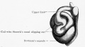
 Horseshoe kidney 01.jpg 701 × 600; 68 KB
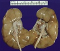
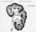
 Horseshoe kidney.jpg 776 × 416; 39 KB
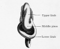
 Horseshoe.jpg 400 × 400; 32 KB
 Human bilateral renal agenesis-hypoplasia-dysplasia.png 1,613 × 906; 2.75 MB
 Human embryonic renal branching 1.jpg 1,280 × 779; 236 KB
 Human embryonic renal branching stage 22.jpg 500 × 756; 150 KB
 Human fetal kidney histology 01.jpg 1,280 × 1,024; 481 KB
 Human fetal kidney histology 02.jpg 1,280 × 1,024; 322 KB
 Human fetal kidney histology 03.jpg 1,280 × 1,024; 333 KB
 Human fetal kidney histology 04.jpg 1,280 × 1,024; 307 KB
 Human- fetal week 10 lower body A.jpg 600 × 450; 96 KB
 Human- fetal week 10 lower body B.jpg 600 × 450; 93 KB
 Human- fetal week 10 lower body C.jpg 600 × 450; 94 KB
 Human- fetal week 10 urogenital A.jpg 600 × 450; 109 KB
 Human- fetal week 10 urogenital B.jpg 600 × 450; 109 KB
 Human- fetal week 10 urogenital C.jpg 600 × 450; 105 KB
 Human- fetal week 10 urogenital D.jpg 600 × 450; 101 KB
 Hydrocolpos.jpg 375 × 361; 23 KB
 Hydronephrosis.jpg 600 × 345; 64 KB
 In vitro kidney development.png 600 × 412; 1.08 MB
 Keibel Mall 2 416.jpg 1,000 × 738; 77 KB
 Keibel Mall 2 520.jpg 1,280 × 998; 167 KB
 Keibel Mall 2 523.jpg 1,280 × 1,234; 208 KB
 Keibel Mall 2 524.jpg 1,280 × 1,072; 208 KB
 Keibel Mall 2 525.jpg 1,280 × 896; 160 KB
 Keibel Mall 2 526.jpg 1,280 × 1,391; 186 KB
 Keibel Mall 2 528.jpg 1,280 × 1,014; 214 KB
 Keibel Mall 2 529.jpg 1,280 × 1,223; 293 KB
 Keibel Mall 2 548.jpg 672 × 483; 53 KB
 Keibel Mall 2 552.jpg 1,280 × 948; 257 KB
 Keibel Mall 2 562.jpg 1,280 × 1,450; 510 KB
 Keibel Mall 2 571.jpg 1,280 × 820; 172 KB
 Keibel Mall 2 572.jpg 1,280 × 969; 219 KB
 Keibel Mall 2 573.jpg 1,280 × 886; 165 KB
 Keibel Mall 2 574.jpg 1,280 × 1,902; 327 KB
 Keibel Mall 2 575.jpg 1,000 × 1,288; 120 KB
 Keibel Mall 2 576.jpg 1,280 × 767; 154 KB
 Keibel Mall 2 578.jpg 1,280 × 836; 115 KB
 Keibel Mall 2 579.jpg 800 × 299; 25 KB
 Keibel Mall 2 583.jpg 1,280 × 1,044; 259 KB
 Keibel Mall 2 584.jpg 464 × 800; 68 KB
 Keibel Mall 2 585.jpg 1,280 × 1,144; 277 KB
 Keibel Mall 2 590.jpg 853 × 702; 66 KB
 Keibel Mall 2 591.jpg 1,004 × 788; 90 KB
 Keibel Mall 2 592.jpg 1,000 × 554; 75 KB
 Keibel Mall 2 596.jpg 1,280 × 1,074; 120 KB
 Keibel Mall 2 598.jpg 1,280 × 854; 146 KB
 Keibel Mall 2 652.jpg 1,200 × 1,036; 130 KB
 Keibel Mall 2 658a.jpg 1,127 × 1,200; 103 KB
 Keibel Mall 2 658b.jpg 895 × 1,200; 98 KB
 Keibel Mall 2 658c.jpg 1,000 × 1,019; 90 KB
 Keibel Mall 2 Felix-plate01.jpg 1,280 × 632; 320 KB
 Keith1902 fig079.jpg 742 × 800; 78 KB
 Keith1902 fig080.jpg 651 × 700; 79 KB
 Keith1902 fig081.jpg 818 × 800; 113 KB
 Keith1902 fig082.jpg 924 × 800; 98 KB
 Keith1902 fig083.jpg 782 × 700; 79 KB
 Keith1902 fig084.jpg 732 × 800; 88 KB
 Keith1902 fig085.jpg 800 × 590; 78 KB
 Keith1902 fig086.jpg 842 × 700; 84 KB
 Keith1902 fig087.jpg 800 × 613; 90 KB
 Keith1902 fig088.jpg 788 × 1,000; 92 KB
 Keith1902 fig089.jpg 964 × 800; 92 KB
 Keith1902 fig090.jpg 700 × 450; 58 KB
 Keith1902 fig091.jpg 700 × 423; 48 KB
 Keith1902 fig093.jpg 700 × 574; 59 KB
 Keith1902 fig094.jpg 800 × 646; 102 KB
 Keith1902 fig095.jpg 660 × 1,000; 125 KB
 Keith1902 fig096.jpg 700 × 535; 69 KB
 Keith1902 fig097.jpg 680 × 592; 80 KB
 Keith1902 fig098.jpg 560 × 460; 40 KB
 Keith1902 fig099.jpg 784 × 800; 105 KB
 Keith1902 fig100.jpg 1,000 × 569; 81 KB
 Keith1902 fig101.jpg 670 × 545; 69 KB
 Keith1902 fig102.jpg 800 × 605; 68 KB
 Keith1902 fig103.jpg 1,000 × 723; 139 KB
 Keith1902 fig104.jpg 800 × 601; 77 KB
 Keith1902 fig105.jpg 632 × 700; 66 KB
 Keith1902 fig108.jpg 780 × 585; 72 KB
 Keith1902 fig109.jpg 859 × 800; 104 KB
 Keith1902 fig110.jpg 800 × 638; 102 KB
 Keith1902 fig111.jpg 1,000 × 620; 145 KB
 Keith1902 fig112.jpg 600 × 541; 77 KB
 Kollmann426.jpg 500 × 620; 46 KB
 Kollmann554.jpg 625 × 568; 54 KB
 Male histology 003.jpg 1,280 × 1,024; 703 KB
 Male histology 004.jpg 1,280 × 1,024; 540 KB
 Male vas deferens and bladder week6to10.jpg 800 × 918; 86 KB
 Mesonephric duct position week 6-11.jpg 697 × 800; 72 KB
 Mouse Kidney Development Cartoon.jpg 506 × 658; 120 KB
 Mouse nephron stages 01.jpg 601 × 916; 217 KB
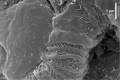
 Mouse renal podocyte EM01.jpg 1,000 × 1,338; 366 KB
 Mouse renal podocyte EM02.jpg 1,000 × 666; 155 KB
 Mouse-kidney in vitro.jpg 955 × 461; 57 KB
 Mouse-kidney model GDNF-FGF10.jpg 405 × 928; 59 KB
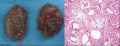 Multicystic kidney and histology.jpg 1,000 × 384; 108 KB
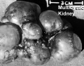 Multicystic kidney.jpg 600 × 468; 56 KB
 Multiple renal arteries 01.jpg 496 × 496; 40 KB
 Neonatal duplicated bladder MRI 01.jpg 751 × 800; 116 KB
 Nephron development 01.jpg 1,232 × 960; 240 KB
 Nephron EM01.jpg 1,909 × 1,280; 219 KB
 Nephron EM02.jpg 1,271 × 1,280; 210 KB
 Nephron EM11.jpg 2,983 × 2,000; 398 KB
 Nephron histology 01.jpg 400 × 500; 79 KB
 Nephron histology 02.jpg 400 × 500; 77 KB
 Nephron histology 03.jpg 375 × 500; 97 KB
 Nephron histology 04.jpg 375 × 500; 54 KB
 Nephron histology.jpg 400 × 500; 70 KB
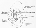
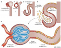
 Nephron physiology.jpg 381 × 262; 49 KB
 Nephrons-cortical and juxtamedullary.jpg 507 × 600; 39 KB


