Thymus Development
Introduction
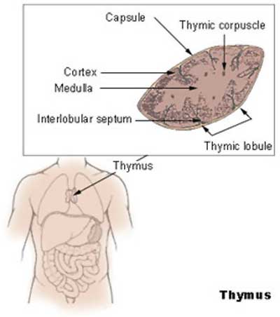
The thymus has a key role in the development of an effective immune system as well as an endocrine function.
The mature thymus epithelium has two main cell types: cortical thymic epithelial (cTECs) and medullary thymic epithelial cells (mTECs) or stromal cells. These thymic stromal cells provide signals for T cell differentiation.
Links: original Endocrine Development - Thymus page
Some Recent Findings
- Rodewald HR. Thymus organogenesis. Annu Rev Immunol. 2008;26:355-88.
- Boehm T, Bleul CC. The evolutionary history of lymphoid organs. Nat Immunol. 2007 Feb;8(2):131-5.
- "During vertebrate evolution, primary lymphoid organs appeared earlier than secondary lymphoid organs. Among the sites of primary lymphopoiesis during evolution and ontogeny, those for B cell differentiation have differed considerably, although they often have had myelolymphatic characteristics. In contrast, only a single site for T cell differentiation has occurred, exclusively the thymus."
- Assarsson E, Chambers BJ, Hogstrand K, Berntman E, Lundmark C, Fedorova L, Imreh S, Grandien A, Cardell S, Rozell B, Ljunggren HG. Severe defect in thymic development in an insertional mutant mouse model. J Immunol. 2007 Apr 15;178(8):5018-27.
Development Overview
The thymus and parathyroid are derived from 3rd pharyngeal pouches.
Development is a series of epithelial/mesenchymal inductive interactions between neural crest-derived arch mesenchyme and pouch endoderm. There is also the possibility that the surface ectoderm of 3rd pharyngeal clefts participates in thymus development.
Hassall's bodies form between 6 and 10 lunar months in humans. They appear after lymphopoiesis has been established and the cortex, medulla and the cortico-medullary junction are able to select of T lymphocytes undergoing progressive maturation. (Text modified from Bodey and Kaiser, 1997)
Experimental studies have shown that a neural crest contribution is also required during early thymic organogenesis.
Cellular and molecular events during early thymus development.[1]
- "The thymic stromal compartment consists of several cell types that collectively enable the attraction, survival, expansion, migration, and differentiation of T-cell precursors. The thymic epithelial cells constitute the most abundant cell type of the thymic microenvironment and can be differentiated into morphologically, phenotypically, and functionally separate subpopulations of the postnatal thymus. All thymic epithelial cells are derived from the endodermal lining of the third pharyngeal pouch. Very soon after the formation of a thymus primordium and prior to its vascularization, thymic epithelial cells orchestrate the first steps of intrathymic T-cell development, including the attraction of lymphoid precursor cells to the thymic microenvironment. The correct segmentation of pharyngeal epithelial cells and their subsequent crosstalk with cells in the pharyngeal arches are critical prerequisites for the formation of a thymus anlage. Mutations in several transcription factors and their target genes have been informative to detail some of the complex mechanisms that control the development of the thymus anlage."
MBoC Figure 24-6. The development and activation of T and B cells
Figure 24-7. Electron micrographs of nonactivated and activated lymphocytes
Development Changes
Changes with age Overall Size
- birth 10-15 g
- puberty 30-40 g
- after puberty - involution
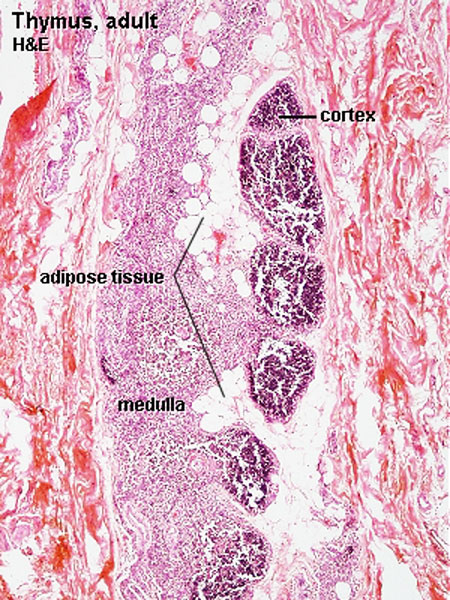
- Replaced by adipose tissue
- middle-aged 10 g
Thymus Anatomy
- Superior mediastinum, anterior to heart
- Bilobed lymphoepithelial organ
- Contains reticular cells but no fibers
- Stem lymphocytes
- proliferate and differentiate
- forms long-lived T- lymphocytes
Thymus Cells
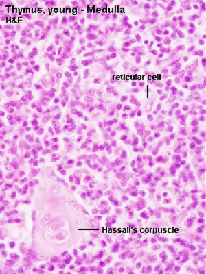
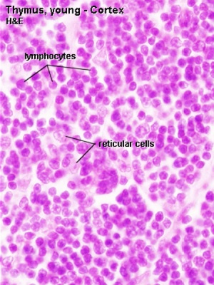
- Reticular cells
- Abundant, eosinophilic, large, ovoid and light nucleus 1-2 nucleoli
- sheathe cortical capillaries
- form an epitheloid layer
- maintain microenvironment for development of T-lymphocytes in cortex (thymic epitheliocytes)
- Macrophages
- cortex and medulla
- difficult to distinguish from reticular cells in H&E
- Lymphocytes
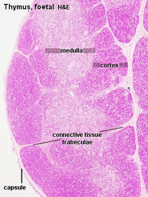
- cortex and medulla - more numerous (denser) in cortex
- majority of them developing T-lymphocytes (= thymic lymphocytes or thymocytes)
Fetal/Young Thymus
|
|
| Young medulla | Young cortex |
Thymic corpuscle
Hassall’s corpuscle - Mass of concentric epithelioreticular cells
Adult Thymus
- Cortical lymphoid tissue is replaced by adipose tissue
- Increase in size of thymic corpuscles
Links: Blue Histology - Thymus
Hassall's Bodies
Hassall's bodies, also called Hassall's corpuscles, form between 6 and 10 lunar months in humans. They appear after lymphopoiesis has been established and the cortex, medulla and the cortico-medullary junction are able to select of T lymphocytes undergoing progressive maturation.Within the thymus their number increases until puberty, then decreases.
Named after Arthur Hill Hassall (1817-1894) a British physician and chemist.
Molecular Development
Both Eva and Six have been implicated in thymus development.[2]
- Eya - human homolog of the Drosophila 'eyes absent' (Eya) gene.
- Six - vertebrate genes which are homologs of the Drosophila 'sine oculis' (so) gene.
Xu PX, Zheng W, Laclef C, Maire P, Maas RL, Peters H, Xu X. Eya1 is required for the morphogenesis of mammalian thymus, parathyroid and thyroid. Development. 2002 Jul;129(13):3033-44.
References
- ↑ Cellular and molecular events during early thymus development. Holländer G, Gill J, Zuklys S, Iwanami N, Liu C, Takahama Y. Immunol Rev. 2006 Feb;209:28-46. Review. PMID: 16448532
- ↑ Patterning of the third pharyngeal pouch into thymus/parathyroid by Six and Eya1. Zou D, Silvius D, Davenport J, Grifone R, Maire P, Xu PX. Dev Biol. 2006 May 15;293(2):499-512. Epub 2006 Mar 10. PMID: 16530750
Articles
- The development of the fetal thymus: an in utero sonographic evaluation. Zalel Y, Gamzu R, Mashiach S, Achiron R. Prenat Diagn. 2002 Feb;22(2):114-7. PMID: 11857615
Search PubMed: Thymus Development
Glossary Links
- Glossary: A | B | C | D | E | F | G | H | I | J | K | L | M | N | O | P | Q | R | S | T | U | V | W | X | Y | Z | Numbers | Symbols | Term Link
Cite this page: Hill, M.A. (2024, May 23) Embryology Thymus Development. Retrieved from https://embryology.med.unsw.edu.au/embryology/index.php/Thymus_Development
- © Dr Mark Hill 2024, UNSW Embryology ISBN: 978 0 7334 2609 4 - UNSW CRICOS Provider Code No. 00098G
