File:Brain-tract-development-03.jpg
Original file (1,000 × 424 pixels, file size: 31 KB, MIME type: image/jpeg)
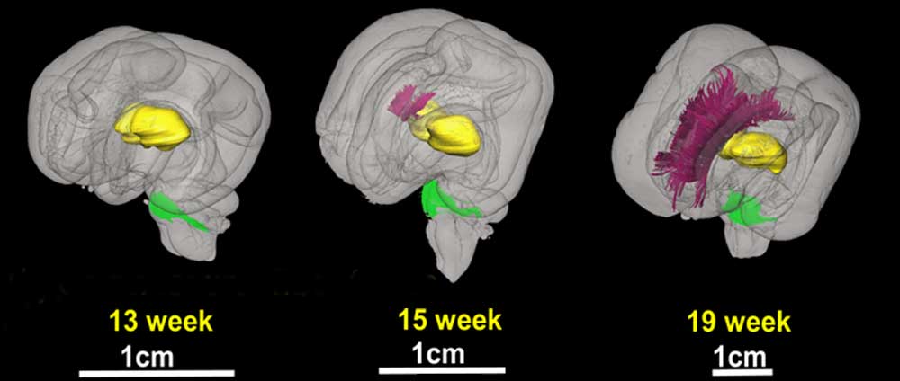
Brain Tract Development - Commissural Fibers
3D depiction of developmental white matter fibers
b is an oblique view of the commissural fibers, where pink and green fibers in 13, 15, and 19 week brains are the corpus callosum and the middle cerebellar peduncle, respectively.
For anatomical guidance, the thalamus (yellow structure) and the ventricles (gray structure) are also shown.
Original File Name: Figure 3 - 3D depiction of developmental white matter fibers.
Reference
<pubmed>19339620</pubmed>| PMC2721010 | J Neurosci.
Copyright: Copyright of all material published in The Journal of Neuroscience remains with the authors. The authors grant the Society for Neuroscience an exclusive license to publish their work for the first 6 months. After 6 months the work becomes available to the public to copy, distribute, or display under a Creative Commons Attribution-Noncommercial-Share Alike 3.0 Unported license.
File history
Yi efo/eka'e gwa ebo wo le nyangagi wuncin ye kamina wunga tinya nan
| Gwalagizhi | Nyangagi | Dimensions | User | Comment | |
|---|---|---|---|---|---|
| current | 12:12, 27 August 2010 | | 1,000 × 424 (31 KB) | S8600021 (talk | contribs) | ==Brain Tract Development - Commissural Fibers== 3D depiction of developmental white matter fibers '''b''' is an oblique view of the commissural fibers, where pink and green fibers in 13, 15, and 19 week brains are the corpus callosum and the middle cer |
You cannot overwrite this file.
File usage
The following page uses this file:


