Category:Cranial Nerve
From Embryology
This Embryology category shows content related to the cranial nerve development (cerebral nerves) that are associated with the brain and the brainstem.
These nerves arise from ectoderm forming both neural crest and epibranchial placodes.
- CN I – Olfactory nerve
- CN II – Optic nerve
- CN III – Oculomotor nerve
- CN IV – Trochlear nerve
- CN V – Trigeminal nerve
- CN VI – Abducens nerve
- CN VII – Facial nerve
- CN VIII – Vestibulocochlear nerve
- CN IX – Glossopharyngeal nerve
- CN X – Vagus nerve
- CN XI – Accessory nerve
- CN XII – Hypoglossal nerve
- Links: cranial nerve | neural
| Neural Crest Links: neural crest | Lecture - Early Neural | Lecture - Neural Crest Development | Lecture Movie | Schwann cell | adrenal | melanocyte | peripheral nervous system | enteric nervous system | cornea | cranial nerve neural crest | head | skull | cardiac neural crest | Nicole Le Douarin | Neural Crest Movies | neural crest abnormalities | Category:Neural Crest | |||
|
Pages in category 'Cranial Nerve'
The following 41 pages are in this category, out of 41 total.
C
- Template:CN I
- Template:CN II
- Template:CN III
- Template:CN IV
- Template:CN IX
- Template:CN V
- Template:CN VI
- Template:CN VII
- Template:CN VIII
- Template:CN X
- Template:CN XI
- Template:CN XII
- Template:Cranial nerve
- Template:Cranial nerve neural crest
- Template:Cranial Nerve Table
- Template:Cranial Nerve Table collapsible
- Template:Cranial Neural Crest Timeline table
P
- Paper - Problems concerning the origin and development of the neural crest and cranial ganglia in the vertebrates (1928)
- Paper - The developing third nerve nucleus in human embryos
- Paper - The Development of the Cranial and Spinal Nerves in the Occipital Region of the Human Embryo
- Paper - The development of the hypoglossal ganglia of pig embryos
- Paper - The hypoglossal nerve in human embryos (1939)
- Paper - The Nuclei of Origin of the Cranial Nerves in the 10 mm Human Embryo
- Paper - The Peripheral Nervous System in the Human Embryo at the End of the First Month (10 mm)
- Paper - The roots of the facial nerve in human embryos and fetuses
- Paper - The spinal accessory nerve in human embryos (1938)
- Paper - The structure of the third, fourth, fifth, sixth, ninth, eleventh and twelfth cranial nerves (1916)
- Paper - The trochlear nerve in human fetuses (1943)
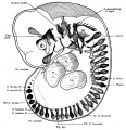
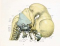
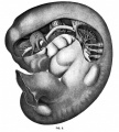
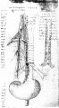
Media in category 'Cranial Nerve'
The following 33 files are in this category, out of 33 total.
 Bailey361.jpg 815 × 662; 86 KB
 Bailey368.jpg 1,074 × 523; 134 KB
 Bailey369.jpg 529 × 446; 33 KB
 Bailey390.jpg 583 × 667; 50 KB
 Bailey402.jpg 640 × 483; 116 KB
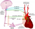 Baroreceptor reflex cartoon.jpg 1,200 × 1,012; 182 KB
 Gray0697.jpg 500 × 540; 49 KB
 Gray0698.jpg 500 × 518; 47 KB
 Gray0778.jpg 600 × 630; 101 KB
 Gray0781.jpg 817 × 700; 156 KB
 Gray0784.jpg 851 × 600; 137 KB
 Gray0788.jpg 815 × 750; 91 KB
 Human Stage14-16 CN5-01.jpg 1,028 × 681; 44 KB
 Keibel Mall 2 032.jpg 1,200 × 750; 139 KB
 Keith1902 fig180.jpg 872 × 800; 141 KB
 Kollmann637.jpg 679 × 561; 51 KB
 Kollmann639.jpg 1,068 × 655; 99 KB
 Kollmann647.jpg 897 × 926; 188 KB
 Lewis1920 fig08.jpg 1,000 × 765; 81 KB
 Mall1905 fig3.jpg 902 × 1,000; 155 KB
 McMurrich1930 fig75.jpg 1,280 × 2,307; 389 KB
 Mouse cranial nerve model SHH.jpg 954 × 900; 74 KB
 Mouse E10.5 Nav2 expression.jpg 1,200 × 818; 186 KB
 Neural - cranial nerves.jpg 800 × 447; 72 KB
 Streeter1906 fig01.jpg 1,193 × 1,318; 167 KB
 Streeter1906 fig02.jpg 1,527 × 2,059; 244 KB
 Streeter1906 fig06.jpg 1,089 × 833; 240 KB
 Streeter1906 fig07.jpg 1,254 × 591; 75 KB
 Streeter1908 fig01.jpg 706 × 1,000; 80 KB
 Streeter1908 fig02.jpg 1,000 × 391; 39 KB
 Streeter1921 fig02.jpg 1,286 × 1,000; 138 KB
 Trigeminal artery 01.jpg 947 × 800; 102 KB
 Trigeminal artery 02.jpg 520 × 490; 35 KB


