File:Meninges cartoon.jpg
From Embryology
No higher resolution available.
Meninges_cartoon.jpg (600 × 385 pixels, file size: 38 KB, MIME type: image/jpeg)
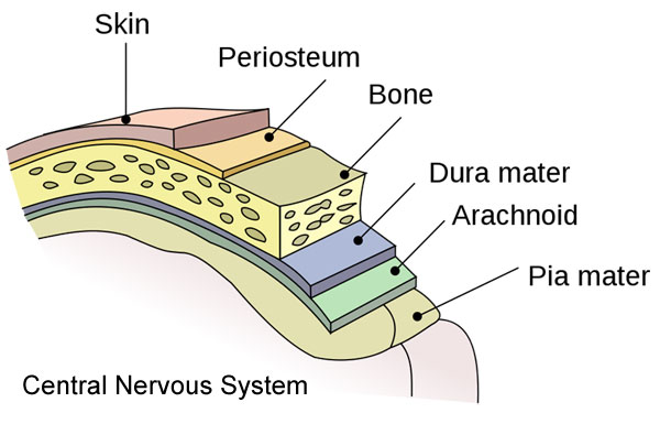
Meninges Diagram
This simplified figure shows the sequence of 3 connective tissue layers that cover the entire central nervous system. The meninges have both neural crest and mesoderm origins.
- Pia mater inner layer lying close the the neural surface.
- Arachnoid mater middle layer containing blood vessels.
- Dura Mater outer layer containing the dural venous sinuses.
- Links: Meninges Development
Reference
Image source: Wikipedia.
Cite this page: Hill, M.A. (2026, October 5) Embryology Meninges cartoon.jpg. Retrieved from https://embryology.med.unsw.edu.au/embryology/index.php/File:Meninges_cartoon.jpg
- © Dr Mark Hill 2026, UNSW Embryology ISBN: 978 0 7334 2609 4 - UNSW CRICOS Provider Code No. 00098G
File history
Yi efo/eka'e gwa ebo wo le nyangagi wuncin ye kamina wunga tinya nan
| Gwalagizhi | Nyangagi | Dimensions | User | Comment | |
|---|---|---|---|---|---|
| current | 11:30, 10 September 2010 | | 600 × 385 (38 KB) | S8600021 (talk | contribs) | ==Meninges diagram== This figure shows the sequence of 3 connective tissue layers that cover the entire central nervous system. # dura mater outer layer # arachnoid middle layer # pia mater inner layer |
You cannot overwrite this file.
File usage
The following 2 pages use this file:


