File:Stage10 neural sm.jpg
Stage10_neural_sm.jpg (665 × 499 pixels, file size: 22 KB, MIME type: image/jpeg)
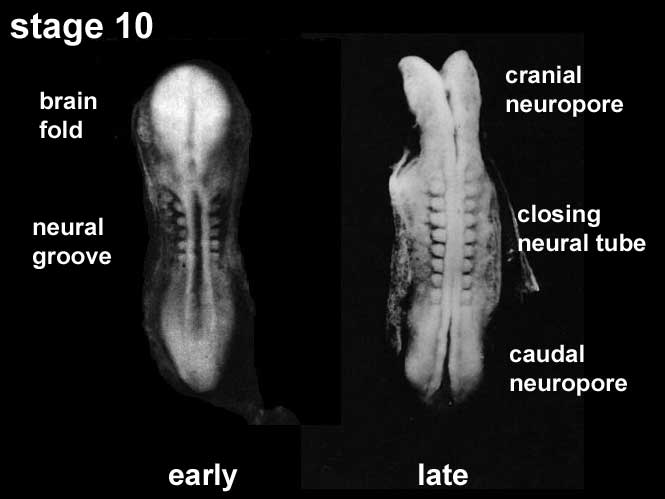
Human Embryo - Neural Plate to Tube
Carnegie stage 10 Week 4 (22 - 23 days) 2 - 3.5 mm, Somite Number 4 - 12
View: This is a dorsal view of the embryo. Top embryo is an early stage 10, bottom is late stage 10. Amniotic membrane removed.
Features: Somite Number 4 - 12, rostral neuropore, neural folds in region of developing brain, neural tube, somites, caudal neuropore, neural fold fuses, remnant of amniotic sac
(note there are 2 separate images of this stage)
Identify: rostral neuropore, neural folds in region of developing brain, neural tube, somites (note the different number formed), caudal neuropore, neural fold fuses, cut edge of amniotic sac
Events Ectoderm: Neural fold deeepens, edges approach midline, neural fold fuses, neural plate folds ventrally in brain region
Mesoderm: Somitogenesis, continued segmentation of paraxial mesoderm (4 - 12 somite pairs)
Cite this page: Hill, M.A. (2026, October 2) Embryology Stage10 neural sm.jpg. Retrieved from https://embryology.med.unsw.edu.au/embryology/index.php/File:Stage10_neural_sm.jpg
- © Dr Mark Hill 2026, UNSW Embryology ISBN: 978 0 7334 2609 4 - UNSW CRICOS Provider Code No. 00098G
File history
Yi efo/eka'e gwa ebo wo le nyangagi wuncin ye kamina wunga tinya nan
| Gwalagizhi | Nyangagi | Dimensions | User | Comment | |
|---|---|---|---|---|---|
| current | 14:28, 10 August 2009 | | 665 × 499 (22 KB) | MarkHill (talk | contribs) | Carnegie stage 10 small image showing neuralation About Carnegie stage 10 Facts: Week 4, 22 - 23 days, 2 - 3.5 mm, Somite Number 4 - 12 View: This is a dorsal view of the embryo. Top embryo is an early stage 10, bottom is late stage 10. Amniotic membra |
You cannot overwrite this file.
File usage
The following 17 pages use this file:
- 2009 Lecture 6
- 2010 BGD Lecture - Development of the Embryo/Fetus 2
- 2010 Lecture 6
- Abnormal Development - Folic Acid and Neural Tube Defects
- BGDA Lecture - Development of the Embryo/Fetus 2
- BGDA Lecture - Development of the Nervous System
- Ectoderm
- Human System Development
- Lecture - Ectoderm Development
- Neural - Amygdala Development
- Neural - Basal Ganglia Development
- Neural - Cerebellum Development
- Neural - Spinal Cord Development
- Neural - Tectum Development
- Neural System - Abnormalities
- Neural System Development
- Talk:BGDA Lecture - Development of the Nervous System


