BGDB Face and Ear - Fetal
Fetal Growth
| <html5media height="480" width="400">File:fetal growth.mp4</html5media>
Click Here to play on mobile device
|
In this cartoon movie of fetal growth, observe the changing relative sizes of the head, body and limbs.
|
Fetal Head Growth

Head Circumference Growth (both gestational and post-fertilisation ages are shown)
Skull Ossification
These are 2 views of the same 12 week 92 mm CRL human fetus head, double stained to show both cartilage (blue) and newly-formed bone (red). The head undergoes two different forms of ossification (endochondral and intramembranous) in separate regions of the skull.
| Lateral view (external) | Medial view (internal) |
|---|---|

|

|
| Note the distribution of new bone formation by intramembranous ossification in the plates of the cranial vault, temporal bone, orbit, upper jaw (maxilla) and lower jaw (mandible) regions. Bony regions in the lower jaw (mandible) region also show spaces where tooth formation is occurring. | Note the distribution of cartilage from the nasal region through the base of the skull showing endochondral ossification, also occuring in the atlas/axis (with new bone forming). See also the original Meckel's cartilage within the newly forming bony mandible. |

Late Fetal Skull
Mandible Ossification


Face
The cartilage template of the mandible and the base of the skull are replaced by early bone development.


Selected midline medial head view showing key features of head musculoskeletal and neurological development.
Note extensive nasal cartilage, nasal conchae, pituitary, secondary palate, oral cavity, tongue, mandible, hyoid, choana, oropharynx.
Also note the developing tongue musculature and its mandibular attachment site.
Note that the cranial vault, the portion of the skull enclosing the brain, ossifies by a unique bone formation process, intramembranous ossification.
Because the head contains many different structures also review notes on Special Senses (eye, ear, nose), Respiration (pharynx), Integumentary (Teeth), Endocrine (thyroid, parathyroid, pituitary) and Musculoskeletal (tongue, skull).
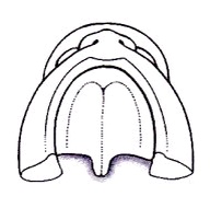
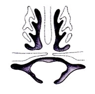
Palate Development

Secondary palate formation is the growth of the palatal shelves towards the midline.
| Inferior view | Anterior view |
|---|---|
| <mediaplayer width='350' height='350' image="http://php.med.unsw.edu.au/embryology/images/4/4f/Palate_001_icon.jpg">File:Palate_001.mp4</mediaplayer> | <mediaplayer width='350' height='350' image="http://php.med.unsw.edu.au/embryology/images/a/a3/Palate_002_icon.jpg">File:Palate_002.mp4</mediaplayer> |
|
|
Palate Overview
Embryonic
Fetal
|
Week 10 Gestational Age (GA week 12)

hard palate |

soft palate |
Hearing

- Week 9 - Mesenchyme surrounding membranous labryinth (otic capsule) chondrifies.
- Week 12-16 - Capsule adjacent to membranous labryinth undegoes vacuolization to form a cavity (perilymphatic space) around membranous labrynth and fills with perilymph.
- Week 18 - ectodermal plug in external auditory meatus breaks down.
- Week 16-24 - Centres of ossification appear in remaining cartilage of otic capsule form petrous portion of temporal bone. Continues to ossify to form mastoid process of temporal bone.
- 3rd Trimester - Vibration acoustically of maternal abdominal wall induces startle response in fetus.

Organ of Corti (mouse, adult)
Central Pathway
- 26 weeks - human brainstem auditory pathway is anatomically formed.
- 28 weeks - AABR can be recorded.
less than 34 weeks - latencies of AABR components (I, III, and V) decrease as a function of gestation
Additional Information
| Additional Information - Content shown under this heading is not part of the material covered in this class. It is provided for those students who would like to know about some concepts or current research in topics related to the current class page. |

Fetal Auricle Development
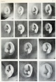
Month 3 - Fetus
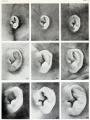
Month 4 - Fetus
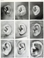
Month 5 - Fetus
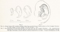
Embryo ear cartilage 21 - 50 mmm CRL


BGDB: Lecture - Gastrointestinal System | Practical - Gastrointestinal System | Lecture - Face and Ear | Practical - Face and Ear | Lecture - Endocrine | Lecture - Sexual Differentiation | Practical - Sexual Differentiation | Tutorial
Glossary Links
- Glossary: A | B | C | D | E | F | G | H | I | J | K | L | M | N | O | P | Q | R | S | T | U | V | W | X | Y | Z | Numbers | Symbols | Term Link
Cite this page: Hill, M.A. (2024, June 21) Embryology BGDB Face and Ear - Fetal. Retrieved from https://embryology.med.unsw.edu.au/embryology/index.php/BGDB_Face_and_Ear_-_Fetal
- © Dr Mark Hill 2024, UNSW Embryology ISBN: 978 0 7334 2609 4 - UNSW CRICOS Provider Code No. 00098G