Category:Ultrasound
From Embryology
This page lists UNSW Embryology content related to ultrasound imaging during development.
Pages in category 'Ultrasound'
The following 38 pages are in this category, out of 38 total.
U
- Ultrasound
- Template:Ultrasound
- Ultrasound - Abnormal
- Ultrasound - Cleft Lip Movie 1
- Ultrasound - Cleft Lip Movie 2
- Ultrasound - Dandy-Walker Movie 1
- Ultrasound - Ectopic Movie 1
- Ultrasound - Ectopic Movie 2
- Ultrasound - Fetal Heart Rate
- Ultrasound - Fetus Movie 1
- Ultrasound - Fetus Movie 2
- Ultrasound - Nuchal Translucency
- Ultrasound - Placenta Previa Movie 1
- Ultrasound - Rabbit Embryo Movie
- Ultrasound - Spina Bifida Movie 1
- Ultrasound - Transposition Great Arteries Movie 1
- Ultrasound - Vasa Previa Movie 1
- Ultrasound - Velamentous Cord Movie 1
- Template:Ultrasound Movies
- Template:Ultrasound Movies - abnormal
- Template:Ultrasound Movies - normal
- Template:Ultrasound Placenta Previa
- Template:Ultrasound Spina-Bifida
- Template:Ultrasound terms
- Template:Ultrasound Transposition Great Arteries
- Template:Ultrasound Vasa Previa
- Template:Ultrasound Velamentous Cord
Media in category 'Ultrasound'
The following 69 files are in this category, out of 69 total.
 Anencephaly ultrasound.jpg 900 × 658; 108 KB
 Aorta coarctation echocardiogram.jpg 601 × 283; 28 KB
 Cervical ectopic ultrasound.jpg 800 × 541; 62 KB
 Cleft lip 01.jpg 585 × 438; 34 KB
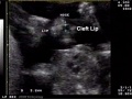 Cleft lip 03.jpg 585 × 439; 38 KB
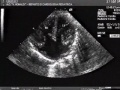 Complete atrioventricular canal.jpg 320 × 240; 22 KB
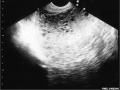 Complete hydatidiform mole 01.jpg 748 × 560; 50 KB
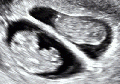 Dichorionic twins ultrasound 01.gif 401 × 282; 2.51 MB
 Echidna egg ultrasound.jpg 484 × 390; 83 KB
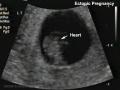 Ectopic 01 heart.jpg 655 × 491; 34 KB
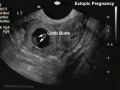 Ectopic 01 limbs.jpg 655 × 491; 37 KB
 Ectopic 01 zoom.jpg 655 × 491; 42 KB
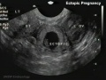 Ectopic 01.jpg 655 × 491; 40 KB
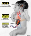 Fetal blood flow liver and brain.jpg 677 × 790; 69 KB
 Fetal cardiac ultrasound 01.jpg 900 × 600; 88 KB
 Fetal ductus venosus pressure wave 01.jpg 706 × 755; 52 KB
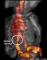 Fetal ductus venosus ultrasound 01.jpg 783 × 1,000; 68 KB
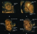 Fetal facial expression 01.jpg 1,200 × 1,094; 122 KB
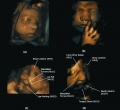 Fetal facial expression 02.jpg 1,914 × 1,762; 205 KB
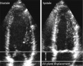 Fetal heart atrioventricular plane displacement 01.jpg 915 × 733; 55 KB
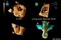 Fetal ultrasound ductal arch 01.jpg 800 × 533; 27 KB
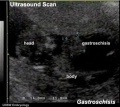 Gastroschisis 01.jpg 493 × 439; 23 KB
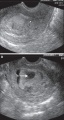 Hydatidiform mole 02.jpg 585 × 1,100; 237 KB
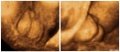 Hypospadia 3D ultrasound 01.jpg 1,150 × 497; 87 KB
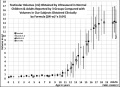 Male puberty testicular volume graph.jpg 1,140 × 826; 126 KB
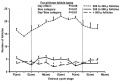 Mouse ovarian follicle size.jpg 600 × 407; 36 KB
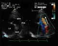 Patent ductus arteriosus echocardiogram.jpg 1,200 × 960; 133 KB
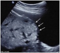 Placenta accreta ultrasound bladder wall interface.jpg 600 × 537; 48 KB
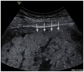 Placenta accreta ultrasound retroplacental clear space loss.jpg 600 × 510; 43 KB
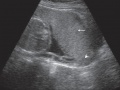 Placenta previa - anterior.jpg 730 × 547; 107 KB
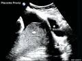 Placenta previa ultrasound 01.jpg 739 × 554; 68 KB
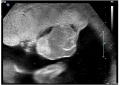 Placental chorioangioma ultrasound 01.jpg 1,183 × 837; 101 KB
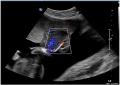 Placental chorioangioma ultrasound 02.jpg 1,183 × 837; 92 KB
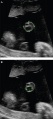 Placental cord ultrasound 01.jpg 585 × 1,309; 192 KB
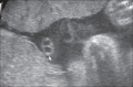 Placental cord ultrasound 02.jpg 731 × 479; 101 KB
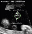 Placental cord ultrasound 03.jpg 598 × 646; 61 KB
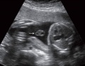 Placental cord ultrasound 04.jpg 729 × 572; 65 KB
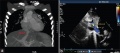 Postnatal persistant ductus venosus ultrasound 01.jpg 1,370 × 600; 96 KB
 Postnatal persistant ductus venosus ultrasound 03.jpg 694 × 600; 54 KB
 Robert Rushmer.jpg 390 × 469; 47 KB
 Septate uterus ultrasound 01.jpg 1,280 × 525; 95 KB
 Tetralogy of Fallot 02.jpg 800 × 796; 63 KB
 Trisomy 10 mosaicism cardiovascular abnormalities.jpg 1,418 × 489; 147 KB
 Trisomy 21 - nuchal translucency graph 01.jpg 638 × 1,000; 69 KB
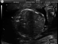 Ultrasound - fetal abdominal circumference.jpg 672 × 512; 23 KB
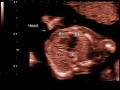 Ultrasound - Hypoplastic left heart syndrome 01.jpg 800 × 600; 53 KB
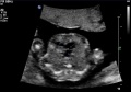 Ultrasound - Hypoplastic left heart syndrome 02.jpg 890 × 626; 54 KB
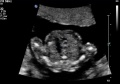 Ultrasound - Hypoplastic left heart syndrome 03.jpg 874 × 612; 51 KB
 Ultrasound - Hypoplastic left heart syndrome 04.jpg 919 × 618; 68 KB
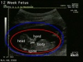 Ultrasound 12 week icon.jpg 500 × 374; 46 KB
 Ultrasound 12wk 4D.mov ; 776 KB
- Ultrasound 12wk 4Dv2.mov ; 1.46 MB
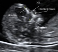 Ultrasound nuchal translucency.jpg 668 × 598; 58 KB
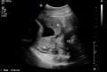 Ultrasound placenta previa 01.jpg 1,024 × 692; 61 KB
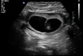 Ultrasound twinning 01.jpg 1,024 × 692; 72 KB
 Ultrasound twinning 02.jpg 1,024 × 692; 71 KB
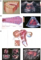 Ultrasound uterine and ovarian vascularity.jpg 531 × 780; 132 KB
 US Circumvallate placenta 01.jpg 1,024 × 692; 61 KB
 US Circumvallate placenta 02.jpg 1,024 × 692; 72 KB
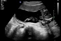 Velamentous cord insertion 01.jpg 1,024 × 692; 78 KB
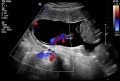 Velamentous cord insertion 02.jpg 1,024 × 692; 104 KB
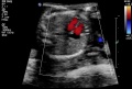 Ventricular septal defect 01.jpg 1,024 × 692; 84 KB
 Z3DCleft Lip Picture.jpg 640 × 480; 32 KB
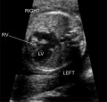 ZPulmonary Atresia.jpg 653 × 618; 85 KB
 ZUltrasound Exam.JPG 1,600 × 1,200; 846 KB
 ZUltrasound Image of Fetal Aorta.jpg 640 × 480; 47 KB
 ZUltrasound machine.jpg 480 × 640; 93 KB
 ZUltrasound System.jpg 262 × 400; 49 KB