File:Adult cochlea cartoon 01.jpg

Original file (986 × 800 pixels, file size: 123 KB, MIME type: image/jpeg)
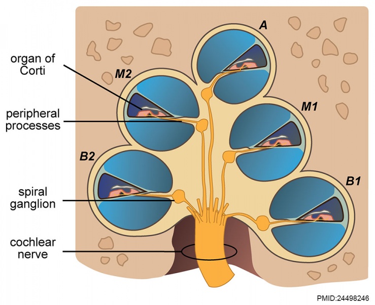
Adult Human Cochlea cartoon
Schematic illustration of a mid-modiolar cut of the adult human cochlea.
- B1 - lower basal turn
- B2 - upper basal turn
- M1 - lower middle turn
- M2 - upper middle turn
- A - apex
- Links: Inner Ear Development | Hearing
Reference
<pubmed>24498246</pubmed>| PLoS One.
Copyright
© 2014 Locher et al. This is an open-access article distributed under the terms of the Creative Commons Attribution License, which permits unrestricted use, distribution, and reproduction in any medium, provided the original author and source are credited.
Locher H, de Groot JCMJ, van Iperen L, Huisman MA, Frijns JHM, et al. (2014) Distribution and Development of Peripheral Glial Cells in the Human Fetal Cochlea. PLoS ONE 9(1): e88066. doi:10.1371/journal.pone.0088066
Image: Figure 1. Capturing PGCs in the human cochlea. doi:10.1371/journal.pone.0088066.g001
Panel B cropped, resized and relabelled from original figure.
File history
Yi efo/eka'e gwa ebo wo le nyangagi wuncin ye kamina wunga tinya nan
| Gwalagizhi | Nyangagi | Dimensions | User | Comment | |
|---|---|---|---|---|---|
| current | 12:33, 7 February 2014 |  | 986 × 800 (123 KB) | Z8600021 (talk | contribs) |
You cannot overwrite this file.
File usage
The following 2 pages use this file: