2009 Lecture 18
Integumentary System
Introduction

The skin provides a barrier between ourselves and our environment, it also contains specializations in different regions including hair, nails, glands and sensory receptors.
The two major tissue organizations of epithelial (ectoderm, epidermis) and mesenchyme (mesoderm connective tissue, dermis and hypodermis) are shown within skin. In addition, we have aslo extensive populating by melanocytes (neural crest) and sensory nerve endings.
It remains today as possibly the first epithelial specialization from which arose other epithelial specializations now located inside the body. The external skin specializations have many different roles and functions. This system is also an excellent model for distribution or "pattern" that is of interest.
Textbooks
- Human Embryology Larson Chapter 14 p443-455
- The Developing Human: Clinically Oriented Embryology (6th ed.) Moore and Persaud Chapter 20: P513-529
Objectives
- Understand the embryonic origin and differentiation of the epidermis and dermis.
- Understand the formation of hair and nails.
- Understand the formation of sweat glands, mammary glands.
- Understand the formation of teeth.
- Brief understanding of associated abnormalities.
Skin Origins
- Ectoderm forms the surface epidermis and the associated glands.
- Mesoderm forms the underlying connective tissue of dermis and hypodermis.
- Neural crest cells also migrate into the forming epidermis and the skin is also populated by specialized sensory endings.
Development Overview
- Ectoderm and Mesoderm Origin
4 weeks
- simple ectoderm epithelium over mesenchyme.
1-3 months
- ectoderm- germinative (basal) cell repeated division of generates stratified epithelium.
- mesoderm- differentiates into connective tissue and blood vessels.
4 months
- basal cell- proliferation generates folds in basement membrane.
- neural crest cells- (melanocytes) migrate into epithelium. These are the pigment cell of the skin.
- embryonic connective tissue- differentiates into dermis, a loose ct layer over a dense ct layer. Beneath the dense ct layer is another loose ct layer that will form the subcutaneous layer.
- Ectoderm contributes to nails, hair follictles and glands.
- Nails form as thickening of ectoderm epidermis at the tips of fingers and toes. These form germinative cells of nail field.
- Cords of these cells extend into mesoderm forming epithelial columns. These form hair follocles, sebaceous and sweat glands.
5 months
- Hair growth initiated at base of cord, lateral outgrowths form associated sebaceous glands.
- Other cords elongate and coil to form sweat glands.
- Cords in mammary region branch as they elongate to form mammary glands. These glands will complete development in females at puberty. Functional maturity only occurs in late pregnancy.
Development of Skin
Skin is our largest organ, providing a protective layer between us and our environment
- epidermis - surface ectoderm
- dermis - mesoderm
- ectoderm/msoderm interaction an inductive manner
- epithelia/mesenchyme (last weeks lecture on kidney)
Regional Specializations
- skin has different structures associated with different regions of the body
- nails, hail, glands, teeth, eyelashes, eyebrow
- 2 main types of histological skin - thin (most of body) thick (soles of feet and hands)
- not the thickest skin
Epidermis
- initially periderm
- keratinization and desquamification
- replaced by basal cells
Epidermis
- week 4-5 early skin is a single ectodermal layer, stratum germinativum basal layer
- week 11 forms intermediate layer
- periderm then lost replaced by stratum corneum, keratinization and desquamification
- week 10 epidermial ridges are formed by proliferation
Neural crest cells
- Neural crest cells migrate into skin (late embryonic) form melanoblasts
- day 40-50 differentiate into then melanocytes - form pigment granules
- different content of melanin (Greek, melas = "black") accounts for differet skin colour
Dermis
- lateral plate mesodermal in origin
- forms connective tissue
- afferent nerves influence dermal ridge formation
Blood Vessels
- lateral plate mesodermal in origin
- week 5 blood vessels form in mesenchyme
- for capillary beds, extensive remodelling with development
Skin Dermatomes
- pattern of skin innervation
- area supplied by single spinal nerve
- motor and sensory DRG
- cutaneous nerve area
Keratin
- large family of intermediate filament protein, 17+ isoforms
- skin disease associated with mutations in keratin genes
Keratins are the major structural proteins of the vertebrate epidermis and its appendages, constituting up to 85% of a fully differentiated keratinocyte. Together with actin microfilaments and microtubules, keratin filaments make up the cytoskeletons of vertebrate epithelial cells. Traced as far back in the evolutionary kingdom as mollusks, keratins belong to the superfamily of intermediate filament (IF) proteins that form alpha-helical coiled-coil dimers which associate laterally and end-to-end to form 10-nm diameter filaments. The evolutionary transition between organisms bearing an exoskeleton and those with an endoskeleton seemed to cause considerable change in keratin. Keratins expanded from a single gene to a multigene family. Of the approximately 60 IF genes in the human genome, half encode keratins, and at least 18 of these are expressed in skin. Vertebrate keratins are subdivided into two sequence types (I and II) that are typically coexpressed as specific pairs with complex expression patterns. The filament-forming capacity of a pair is dependent upon its intrinsic ability to self-assemble into coiled-coil heterodimers, a feature not required of the invertebrate keratins (Weber et al 1988). Approximately 20,000 heterodimers of type I and type II keratins assemble into an IF. Mutations that perturb keratin filament assembly in vitro can cause blistering human skin disorders in vivo. (from Review Article)
Development of Glands
- 2 main types - sebaceous and sweat
- both ectodermal in origin
- form as ingrowth of ectoderm into the mesoderm
Sebaceous
- associated with hair development
- except plans penis and labia minora
- these glands secrete vernix
Sweat Glands
- mostly eccrine some apocrine
- apocrine in axilla, pubic and nipple regions
- see also mammary gland development
Vernix Caseosa
- (Latin, vernix caseosa = varnish)
- covers fetal skin- secretion from sebaceous glands
- protects skin from extraembryonic fluids amnion, urine
- slippery and helps with parturition
Hair Development
- week 9 -12
- follicle forms in st. germinativum of epidermis
- hair bud -> hair bulb
- hair bulb forms hair
- mesenchyme forms hair papilla
- germinal matrix cells become keratinized to form hair shaft
- arrector pili muscle develop in mesenchyme
- week 12 - lanugo hair (L. lana = wool)
- first hair formed replaced postnatally
- bind vernix to skin
- Melanocytes (neural crest) produce melanin which influences hair colour
Postnatal Development
- puberty - coarse hair in pubis and axilla
- males face, chest, etc
Molecular Hair Development
- EGF inhibits hair follicle formation
- SHH Knockout mice
- Hair germs comprising epidermal placodes and associated dermal condensates were detected in both control and Shh -/- embryos, but progression through subsequent stages of follicle development was blocked in mutant skin. The expression of Gli1 and Ptc1 was reduced in Shh -/- dermal condensates and they failed to evolve into hair follicle papillae, suggesting that the adjacent mesenchyme is a critical target for placode-derived Shh. Despite the profound inhibition of hair follicle morphogenesis, late-stage follicle differentiation markers were detected in Shh -/- skin grafts, as well as cultured vibrissa explants treated with cyclopamine to block Shh signaling. Our findings reveal an essential role for Shh during hair follicle morphogenesis, where it is required for normal advancement beyond the hair germ stage of development. Chin etal Developmental Biology, v 205, n 1, Jan 1999, p 1-9
Molecular Hair Development
- inappropriate activation of the Shh signal
- shh transduction cascade in human epidermis can cause basal cell carcinoma. Here we show that during normal development of avian skin, Shh is first expressed only after the responsiveness to this protein has been suppressed in most of the surrounding ectodermal cells. Forced expression of Shh in avian skin prior to this time causes a disorganized ectodermal proliferation. However, as skin begins to differentiate, the forced expression of Shh causes feather bud formation. Subsequently, expression of Shh in interfollicular epidermis has little or no morphological effect. Restricted responsiveness to Shh in developing skin has functional consequences for morphogenesis and may have important implications for cutaneous pathologies as well
- Morgan etal. Developmental Biology, v 201, n 1, September 1, 1998, p1-12
Nail Development
- week 10 fingernails before toenaois
- as in limb development
- nail field
- appears at tip and migrates to dorsal surface
- thickened epidermis
- surrounding cells form nail fold
- keratinization of proximal nail fold forms nail plate
- nails reach tip
- week 32 fingernails
- week 36 toenails
- nail growth indicator of prematurity
Mammary Glands
- week 6 epidermis downgrowth into dermis
- modified sweat glands
- epithelia/mesenchyme inductive interaction
- mesenchyme forms connective tissue and fat
- mammary ridges- mammary bud formation
- pair of ventral regions axilla to inguinal
- pectoral regions generate breasts
- buds branch to form lactiferous ducts
- placental sex hormone
- only main duct formed at birth
- Mammary Glands
- mammary pit
- forms fetal period
- depressed region at gland
- areola
- proliferation of connective tissue postnatally
- prior to puberty male and female glands the same
- Mammary Glands-Puberty
- sex hormone estrogen stimulate growth
- full development approx 20 years
- mainly fat and connective tissue deposition
- growth also influenced by other hormones
- progereterone, prolactin, corticoids, growth hormone
Breast cancer
- In 1994, two breast cancer susceptibility genes were identified: BRCA1 on chromosome 17 BRCA2 on chromosome 13
- When an individual carries a mutation in either BRCA1 or BRCA2, they are at an increased risk of being diagnosed with breast or ovarian cancer at some point in their lives. Normal function of these genes was to participate in repairing radiation-induced breaks in double-stranded DNA. It is though that mutations in BRCA1 or BRCA2 might disable this mechanism, leading to more errors in DNA replication and ultimately to cancerous growth.
Breast Cancer Detection - reduce mortality is through early detection (general screening of the population for BRCA1 and BRCA2 is not yet recommended). New strategies to find anti-cancer drugs are constantly being developed. The latest, called 'synthetic lethal screening' looks for new drug targets in organisms such as yeast and fruit flies. In the same way that studies in yeast recently helped to identify the functions of BRCA1 and BRCA2, it is thought that drugs that work in more primative organisms will also be applicable to humans.
Mammary Glands-Pregnancy
- raised estrogens and progesterone stimulate gland development
- hemispherical shape due to fat deposition
- lactation supports development
Breast Abnormalities
- occurs in 1% of female population
- polymastia - extra breast
- polytheli - extra nipple, supernumerary nipple (relatively common in males)
Glands
Hair
- Before birth we have embryonic hair that has an imporant role in binding the skins waxy protective coating against our watery environment.
- After birth we have early postnatal hair that is gradually replaced by the mature form.
- At puberty we have a second round of hair formation under endocrine regulation by sex hormones.
Fetal Hair
- Month 5 - hair appears on the head and beginning of vernix caseosa deposition.
- Month 6 - body is covered by fine hairs (lanugo) and the deposit of vernix caseosa is considerable. Skin papilla are developed and the free border of the nail projects from the corium of the dermis.
- Month 8 - skin now completely coated with vernix caseosa, and the lanugo begins to disappear. Skin is also pink in colour and subcutaneous fat being deposited (hypodermis layer).
- Month 9 - lanugo has largely disappeared from the trunk.
Teeth
- integumentary system specialization by epitheilal/mesenchymal interactions in development and develops with a major contribution from the neural crest.
- ectoderm of the first pharyngeal arch and neural crest, ectomesenchymal cells
Links
- UNSW Embryology - Introduction | Detailed Overview of Skin components | Abnormalities | Stage 13/14 | Stage 22 | Histology | Dermatomes | Glands | Mammary Glands | Teeth | Hair | Nails
References
- Before We Are Born (5th ed.) Moore and Persaud Chapter 21: P481-496
- Essentials of Human Embryology Larson Chapter 14: P303-315
- Color Atlas of Clinical Embryology Moore Persaud and Shiota Chapter 15: p231-236
- Nature - Milestones in Cutaneous Biology "highlights groundbreaking advances in cutaneous biology over the past 100 years."
- International Journal Developmental Biology 2004 - Skin Development special issue
Images
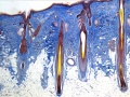
Hair histology
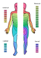
Dermatomes
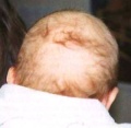
Neonate hair


Glossary Links
- Glossary: A | B | C | D | E | F | G | H | I | J | K | L | M | N | O | P | Q | R | S | T | U | V | W | X | Y | Z | Numbers | Symbols | Term Link
Course Content 2009
Embryology Introduction | Cell Division/Fertilization | Cell Division/Fertilization | Week 1&2 Development | Week 3 Development | Lab 2 | Mesoderm Development | Ectoderm, Early Neural, Neural Crest | Lab 3 | Early Vascular Development | Placenta | Lab 4 | Endoderm, Early Gastrointestinal | Respiratory Development | Lab 5 | Head Development | Neural Crest Development | Lab 6 | Musculoskeletal Development | Limb Development | Lab 7 | Kidney | Genital | Lab 8 | Sensory - Ear | Integumentary | Lab 9 | Sensory - Eye | Endocrine | Lab 10 | Late Vascular Development | Fetal | Lab 11 | Birth, Postnatal | Revision | Lab 12 | Lecture Audio | Course Timetable
Cite this page: Hill, M.A. (2026, October 28) Embryology 2009 Lecture 18. Retrieved from https://embryology.med.unsw.edu.au/embryology/index.php/2009_Lecture_18
- © Dr Mark Hill 2026, UNSW Embryology ISBN: 978 0 7334 2609 4 - UNSW CRICOS Provider Code No. 00098G
