Category:Histology
From Embryology
Introduction
Files and media below relate to histology. Most links are to histological images that relate to tissue structure and development. Many images are sourced from the original UNSW Anatomy Histology slide set and UWA Blue Histology online images.
Subcategories
This category has the following 15 subcategories, out of 15 total.
Pages in category 'Histology'
The following 134 pages are in this category, out of 334 total.
(previous page) (next page)P
- Paper - A morphological study of the development of the human liver 1
- Paper - A morphological study of the development of the human liver 2
- Paper - A note on the post-natal growth of the kidney, thyroid gland and liver (1924)
- Paper - A quantitative study of the fetal growth changes in the parts of the human stomach wall
- Paper - A Study of the Structural Unit of the Liver
- Paper - A subject with complete transposition of viscera (1917)
- Paper - Appendix vermiformis duplex (1936)
- Paper - Case of abnormal duodenum (1924)
- Paper - Changes in fetuses due to formalin preservation
- Paper - Chiefly concerning the genito-mesenteric fold of peritoneum
- Paper - Congenital absence of the appendix of the caecum (1915)
- Paper - Congenital anomalies of the duodenum (1940)
- Paper - Congenital Anomalies of the Liver (1929)
- Paper - Congenital atresia of the oesophagus
- Paper - Congenital hernia into the umbilical cord - two cases, one associated with persistent cloaca
- Paper - Congenital malformations of the oesophagus
- Paper - Cytogenesis of the human fetal pancreas (1962)
- Paper - Cytological studies of Langerhans's islets, with special reference to the problem of their relation to the pancreatic acinus tissue (1920)
- Paper - Early differentiation of the foregut in the dog
- Paper - Imperfect torsion of the intestinal loop
- Paper - Normal development of the trachea and esophagus in man
- Paper - Notes on the origin of the liver (1891)
- Paper - Obstructions about the mesentery in infants (1936)
- Paper - On abnormalities of the caecum and colon with reference to development
- Paper - On the development of the villi of the human intestine
- Paper - On the developmental topography of the thoracic and abdominal viscera (1909)
- Paper - On the factors concerned in causing rotation of the intestine in man
- Paper - On the histogenesis of gastric glands
- Paper - On the relation of the liver cells to the blood-vessels and lymphatics
- Paper - On the so-called ultimobranchial body of the mammalian embryo (1915)
- Paper - Retrogressive Changes in the Fetal Vessels and the Suspensory Ligament of the Liver
- Paper - Sequential innervation of the intestinal loop in the human embryo
- Paper - Some factors influencing the position of the small intestine (1915)
- Paper - Studies of the intestine and peritoneum in the human foetus - part 1
- Paper - Studies of the intestine and peritoneum in the human foetus - part 2
- Paper - Studies of the intestine and peritoneum in the human foetus - part 3
- Paper - Studies of the intestine and peritoneum in the human foetus - part 4
- Paper - Studies of the intestine and peritoneum in the human foetus - part 5
- Paper - Studies of the intestine and peritoneum in the human foetus - part 6
- Paper - The angiology, angiogenesis, and organogenesis of the submaxillary gland
- Paper - The application of trichrome staining methods to embryological technique (1940)
- Paper - The bi-lobed form of the ventral pancreas in mammals
- Paper - The comparative anatomy of the lips and labial villi of vertebrates
- Paper - The critical period in the development of the intestines (1914)
- Paper - The development of the form of the gastrointestinal canal in humans 1
- Paper - The development of the form of the gastrointestinal canal in humans 2
- Paper - The development of the great omentum and transverse mesocolon
- Paper - The development of the human pharynx
- Paper - The development of the lobule of the pig's liver (1919)
- Paper - The development of the lobus quadratus of the liver with special reference to an unusual anomaly of this lobe in the adult (1914)
- Paper - The development of the mucous membrane oesophagus stomach and small intestine in human embryo
- Paper - The development of the mucous membrane of the large intestine and vermiform process in the human embryo
- Paper - The development of the rectum in the human embryo
- Paper - The development of the serous glands (von Ebner's) of the vallate papillae in man (1917)
- Paper - The development of the spiral coil in the large intestine of the pig
- Paper - The early looping of the alimentary canal in the mammalian and human foetus and the mechanisms assumed to be active in this process
- Paper - The early stages of the development of the ileo-colic sphincter (1924)
- Paper - The embryogenesis of human bile capillaries and ducts
- Paper - The form of the stomach in human embryos with notes upon the nomenclature of the stomach
- Paper - The formation of the duodenal curve
- Paper - The formation of the duodenal curve (1919)
- Paper - The gall bladder and the extrahepatic biliary passages in late embryonic and early fetal life
- Paper - The genesis of Jackson's membrane (1914)
- Paper - The genesis of Jackson's membrane: notes on the genito-mesenteric fold of peritoneum and the supra-adhesion foramen
- Paper - The lachrymal gland (1916)
- Paper - The morphology and development of intestinal folds and villi in vertebrates
- Paper - The nature of the malformations of the rectum and urogenital passages
- Paper - The origin of blood cells (1916)
- Paper - The regular occurrence of intestinal diverticula in embryos of the pig, rabbit and man
- Paper - The regular occurrence of intestinal diverticula in embryos of the pig, rabbit, and man
- Paper - The relations of endogenous and exogenous factors in bone and tooth development (1937)
- Paper - The relative frequency of the various positions of the vermiform appendix (1924)
- Paper - The role of the primitive mesothelium in the development of the mammalian spleen (1936)
- Paper - The shrinkage of embryos in the processes preparatory to sectioning
- Paper - Transposition of Abdominal Viscera (1926)
- Paper - V. Meckel's diverticulum patent at the navel (1902)
- Template:Peripheral Nerve Histology
- Placenta - Histology
- Template:Placenta Cord Histology
- Template:Placenta histology
R
- Template:Ref-Arey1917
- Template:Ref-BacsichSmout1938
- Template:Ref-Badertscher1915b
- Template:Ref-Baxter1940
- Template:Ref-Bloom1931
- Template:Ref-BöhmDavidoffHuber1910
- Template:Ref-Cooper1938
- Template:Ref-Danchakoff1916b
- Template:Ref-Danchakoff1916c
- Template:Ref-Danchakoff1918
- Template:Ref-HallpikePeet1939
- Template:Ref-Hassall1849
- Template:Ref-Herring1908a
- Template:Ref-Ingalls1915
- Template:Ref-Lewis1906
- Template:Ref-Nonidez1941
- Template:Ref-Orban1944
- Template:Ref-PattenPhilpott1921
- Template:Ref-ScharpenbergWindle1938
- Template:Ref-Schultz1919
- Template:Ref-ThielDowney1921
- Template:Ref-WheatersHistology2006
- Template:Ref-WislockiDempsey1945
- Template:Renal Histology
- Template:Renal histology
- Renal System Histology
- Respiratory System - Histology
S
- Salivary Gland Development
- Sertoli cell
- SH Lecture - Lymphatic Structure and Organs
- SH Practical - Lymphatic Structure and Organs
- SH Practical - Respiratory
- Site Map
- Talk:Site Map
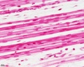
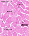
- Skeletal Muscle Histology
- Template:SmMhistolinks
- Smooth Muscle Development
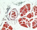
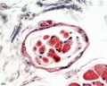
- Smooth Muscle Histology
- Template:Spalteholz method
- Template:Spleen Histology
- Template talk:Spleen Histology
- Template:Spleen Histology gallery
- Template:Stage 22 histology gallery
- Template:Stage 22 histology gallery table
- Template:Stains
- Template:Stomach Histology
T
Media in category 'Histology'
The following 200 files are in this category, out of 718 total.
(previous page) (next page)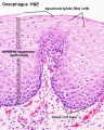 Oesophagus histology 04.jpg 480 × 600; 104 KB
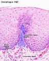 Oesophagus histology 05.jpg 480 × 600; 100 KB
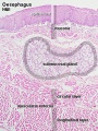 Oesophagus histology 06.jpg 400 × 533; 100 KB
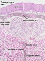 Oesophagus histology 07.jpg 400 × 533; 100 KB
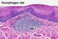 Oesophagus MALT.jpg 500 × 333; 73 KB
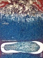
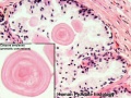
 Ossification centre.jpg 450 × 600; 101 KB
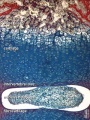
 Ossification endochondral 01.jpg 817 × 613; 198 KB
 Ossification endochondral 1.jpg 750 × 1,000; 147 KB
 Ossification endochondral 1a.jpg 600 × 800; 103 KB
 Ossification endochondral 1b.jpg 450 × 600; 64 KB
 Ossification endochondral 1c.jpg 300 × 400; 32 KB
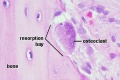 Osteoclast.jpg 500 × 333; 41 KB
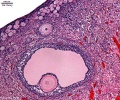
 Ova20he.jpg 450 × 600; 96 KB
 Ova41he.jpg 450 × 600; 113 KB
 Ova44he.jpg 1,280 × 1,024; 315 KB
 Ovary corpus luteum.jpg 2,178 × 1,137; 376 KB
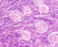
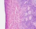
 Ovary Histology - tunica albuginea.jpg 1,280 × 1,024; 336 KB
 Ovary histology 001.jpg 1,280 × 1,024; 360 KB
 Ovary histology 002.jpg 1,280 × 1,024; 270 KB
 Ovary histology 003.jpg 1,280 × 1,024; 337 KB
 Ovary histology 004.jpg 1,280 × 1,024; 401 KB
 Ovary histology 005.jpg 1,280 × 1,024; 354 KB
 Ovary histology 006.jpg 1,280 × 1,024; 424 KB
 Ovary histology 007.jpg 1,280 × 1,024; 336 KB
 Ovary histology 008.jpg 1,280 × 1,024; 264 KB
 Ovary histology 061.jpg 1,280 × 1,024; 438 KB
 Ovary histology 061a.jpg 800 × 640; 200 KB
 Ovary histology 061c.jpg 400 × 320; 56 KB
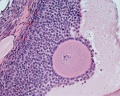
 Ovary- atretic follicle 01.jpg 793 × 595; 225 KB
 Ovary- atretic follicle 02.jpg 600 × 450; 139 KB
 Ovary- atretic follicle 03.jpg 790 × 593; 202 KB
 Ovary- atretic follicle 04.jpg 600 × 450; 128 KB
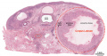
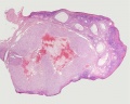
 Ovary- histology overview.jpg 861 × 646; 160 KB
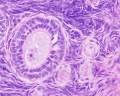
 Ovary- histology secondary follicle 01.jpg 1,000 × 800; 293 KB
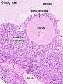
 Ovary10x.jpg 480 × 400; 53 KB
 Ovary5x.gif 480 × 400; 162 KB
 Pacinian corpuscle histology 01.jpg 800 × 640; 227 KB
 Pacinian corpuscle histology 02.jpg 1,280 × 1,024; 207 KB
 Pacinian corpuscle histology 03.jpg 1,280 × 1,024; 133 KB
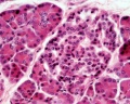
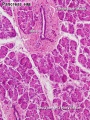 Pancreas histology 001.jpg 375 × 500; 90 KB
 Pancreas histology 002.jpg 375 × 500; 50 KB
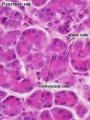
 Pancreas histology 003.jpg 375 × 500; 90 KB
 Pancreas histology 004.jpg 400 × 267; 50 KB
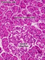
 Pancreas histology 005.jpg 375 × 500; 64 KB
 Pancreas histology 101.jpg 1,280 × 1,024; 505 KB
 Pancreas histology 102.jpg 1,280 × 1,024; 309 KB
 Pancreas histology 103.jpg 1,280 × 1,024; 313 KB
 Pancreas histology 104.jpg 1,280 × 1,024; 371 KB
 Pancreas histology 105.jpg 1,280 × 1,024; 277 KB
 Pancreas histology 106.jpg 1,280 × 1,024; 314 KB
 Pancreas histology 10he.jpg 300 × 400; 73 KB
 Pancreas histology 40he.jpg 300 × 400; 42 KB
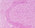
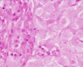
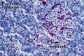
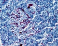
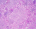
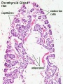 Parathyroid histology 001.jpg 450 × 600; 54 KB
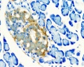
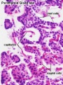 Parathyroid histology 002.jpg 450 × 600; 43 KB
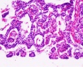
 Parathyroid histology 003.jpg 1,280 × 1,024; 161 KB
 Parathyroid histology 004.jpg 1,280 × 1,024; 162 KB
 Patrick de Permentier.jpg 200 × 200; 12 KB
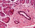
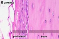 Periosteum.jpg 500 × 333; 34 KB
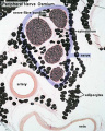
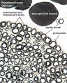
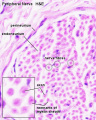 Peripheral nerve histology 01.jpg 640 × 800; 56 KB
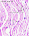 Peripheral nerve histology 02.jpg 640 × 800; 53 KB
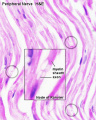 Peripheral nerve histology 03.jpg 640 × 800; 51 KB
 Peripheral nerve histology 04.jpg 640 × 800; 79 KB
 Peripheral nerve histology 05.jpg 640 × 800; 78 KB
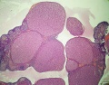
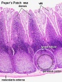 Peyer's patch 01.jpg 450 × 600; 118 KB
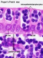 Peyer's patch 02.jpg 450 × 600; 69 KB
 Philipp Stöhr.jpg 637 × 779; 68 KB
 Pineal histology 001.jpg 450 × 600; 75 KB
 Pineal histology 002.jpg 1,000 × 800; 241 KB
 Pineal histology 003.jpg 800 × 640; 166 KB
 Pituitary development animation.gif 600 × 400; 272 KB
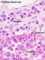 Pituitary histology 001.jpg 450 × 600; 72 KB
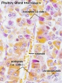 Pituitary histology 002.jpg 450 × 600; 81 KB
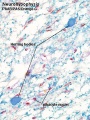 Pituitary histology 003.jpg 450 × 600; 94 KB
 Pituitary histology 004.jpg 1,280 × 1,024; 342 KB
 Pituitary histology 005.jpg 1,280 × 1,024; 326 KB
 Pituitary histology 006.jpg 1,280 × 1,024; 450 KB
 Pituitary histology 007.jpg 1,280 × 1,024; 325 KB
 Pituitary histology 008.jpg 1,280 × 1,024; 340 KB
 Pituitary histology 009.jpg 466 × 610; 64 KB
 Pituitary histology 010.jpg 1,005 × 961; 249 KB
 Pituitary histology 011.jpg 900 × 1,388; 309 KB
 Placenta anchoring villi.jpg 600 × 450; 167 KB
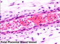 Placenta blood.jpg 450 × 333; 51 KB
 Placenta histology 001.jpg 1,280 × 1,024; 155 KB
 Placenta histology 002.jpg 1,280 × 1,024; 102 KB
 Placenta histology 003.jpg 1,280 × 1,024; 58 KB
 Placenta histology 004.jpg 1,280 × 960; 526 KB
 Placenta histology 005.jpg 1,280 × 960; 433 KB
 Placenta histology 006.jpg 666 × 500; 94 KB
 Placenta histology 007.jpg 1,265 × 437; 207 KB
 Placenta histology 008.jpg 800 × 599; 198 KB
 Placenta Hofbauer cells 01.jpg 934 × 700; 156 KB
 Placenta percreta 01.jpg 1,200 × 904; 327 KB
 Placenta percreta 05.jpg 900 × 676; 77 KB
 Placenta- first trimester histology x40.jpg 1,000 × 800; 124 KB
 Placental artery 01.jpg 1,200 × 838; 371 KB
 Placental artery.jpg 600 × 509; 78 KB
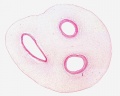
 Placental cord cross-section 01.jpg 1,184 × 1,000; 277 KB
 Placental cord cross-section.jpg 525 × 525; 50 KB
 Placental cord epithelium 01.jpg 1,200 × 805; 130 KB
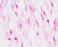
 Placental trophospongium.jpg 567 × 344; 94 KB
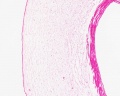
 Placental vein.jpg 812 × 392; 59 KB
 Placental villi 1.jpg 1,280 × 1,024; 77 KB
 Placental villi 2.jpg 1,280 × 1,024; 70 KB
 Placental villi 3.jpg 1,280 × 1,024; 99 KB
 Placental villi 4.jpg 1,280 × 1,024; 89 KB
 Placental villi 5.jpg 1,280 × 1,024; 238 KB
 Placental villi 6.jpg 1,000 × 750; 277 KB
 Placental villi.jpg 1,280 × 1,024; 199 KB
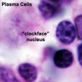 Plasma cell clockface nucleus 01.jpg 400 × 400; 27 KB
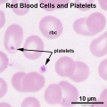 Platelet 01.jpg 600 × 600; 57 KB
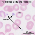 Platelet 02.jpg 600 × 600; 57 KB
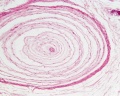
 Proboscis histology.jpg 600 × 1,041; 166 KB
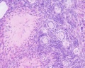
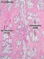 Prostate histology 01.jpg 300 × 400; 72 KB
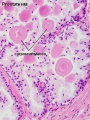 Prostate histology 02.jpg 300 × 400; 57 KB
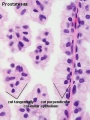 Prostate histology 03.jpg 300 × 400; 41 KB
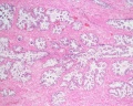 Prostate histology 04.jpg 1,280 × 1,024; 569 KB
 Prostate histology 05.jpg 1,280 × 1,024; 418 KB
 Prostate histology 06.jpg 1,280 × 1,024; 348 KB
 Prostate histology 07.jpg 1,280 × 1,024; 328 KB
 Prostate histology 08.jpg 1,280 × 1,024; 252 KB
 Prostate histology 09.jpg 1,019 × 764; 199 KB
 Rat ovary histology 01.jpg 1,200 × 938; 274 KB
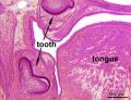 Rat-neonatal teeth.jpg 300 × 230; 38 KB
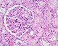
 Renal histology 01.jpg 1,280 × 1,024; 684 KB
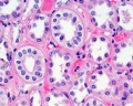
 Renal histology 02.jpg 1,280 × 1,024; 376 KB
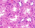
 Renal histology 03.jpg 1,280 × 1,024; 280 KB
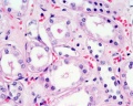
 Renal histology 04.jpg 1,280 × 1,024; 266 KB
 Renal histology 05.jpg 1,280 × 1,024; 275 KB
 Renal histology 06.jpg 1,280 × 1,024; 579 KB
 Renal histology 07.jpg 1,280 × 1,024; 396 KB
 Renal histology 08.jpg 1,280 × 1,024; 293 KB
 Respiratory histology 01.jpg 450 × 600; 86 KB
 Respiratory histology 02.jpg 450 × 600; 37 KB
 Respiratory histology 03.jpg 450 × 600; 29 KB
 Respiratory histology 04.jpg 450 × 600; 31 KB
 Respiratory histology 05.jpg 450 × 600; 96 KB
 Respiratory histology 06.jpg 450 × 600; 95 KB
 Respiratory histology 07.jpg 1,280 × 1,024; 255 KB
 Respiratory histology 08.jpg 1,280 × 1,024; 263 KB
 Respiratory histology 09.jpg 1,280 × 1,024; 236 KB
 Respiratory histology 11.jpg 450 × 600; 65 KB
 Respiratory histology 12.jpg 450 × 600; 88 KB
 Respiratory histology 13.jpg 450 × 600; 102 KB
 Respiratory histology 14.jpg 450 × 600; 87 KB
 Reticulocyte.jpg 500 × 313; 14 KB
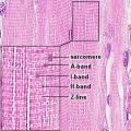
 Sarcomere animation.gif 400 × 200; 115 KB
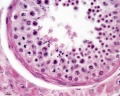
 Seminiferous-tubule-HEx40.jpg 400 × 500; 59 KB
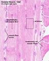
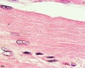
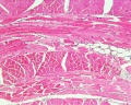 Skeletal muscle histology 001.jpg 1,280 × 1,024; 562 KB
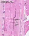
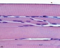
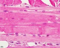 Skeletal muscle histology 002.jpg 1,280 × 1,024; 352 KB
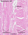
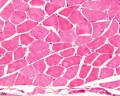 Skeletal muscle histology 003.jpg 1,280 × 1,024; 411 KB
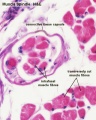
 Skeletal muscle histology 004.jpg 1,280 × 1,024; 242 KB
 Skeletal muscle histology 005.jpg 1,280 × 1,024; 288 KB
 Skeletal muscle histology 006.jpg 1,280 × 1,024; 237 KB
 Skeletal muscle histology 007.jpg 1,280 × 1,024; 253 KB
 Skeletal muscle histology 008.jpg 1,280 × 1,024; 237 KB
 Skeletal muscle histology 009.jpg 1,280 × 1,024; 274 KB
 Skeletal muscle histology 010.jpg 1,280 × 1,024; 207 KB
 Skeletal muscle histology 011.jpg 600 × 750; 132 KB
 Skeletal muscle histology 012.jpg 600 × 750; 121 KB
 Skeletal muscle histology 013.jpg 600 × 750; 185 KB
 Skeletal muscle histology 014.jpg 600 × 750; 127 KB
 Skeletal muscle histology 015.jpg 600 × 750; 84 KB
 Skeletal muscle histology 016.jpg 450 × 450; 92 KB
 Skeletal muscle histology 017.jpg 1,280 × 1,024; 357 KB
 Skeletal muscle histology 018.jpg 1,280 × 1,024; 290 KB
 Skeletal muscle histology 022.jpg 1,280 × 1,024; 471 KB
 Skeletal muscle histology 044.jpg 480 × 600; 74 KB
 Skeletal muscle histology 055.jpg 1,280 × 1,024; 333 KB
 Skeletal muscle histology 077.jpg 1,280 × 1,024; 286 KB
 Skeletal muscle histology 444.jpg 934 × 701; 125 KB
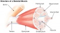 Skeletal muscle structure cartoon.jpg 520 × 286; 46 KB
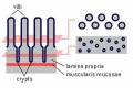 Small intestine villi and crypts.jpg 500 × 333; 26 KB
 Smear- early proliferative.jpg 521 × 350; 47 KB
 Smear- late secretory.jpg 525 × 350; 74 KB
 Smear- late-proliferative.jpg 511 × 350; 46 KB
 Smear- mid-proliferative.jpg 519 × 350; 58 KB
 Smear- secretory.jpg 520 × 350; 57 KB
 Smooth muscle histology 001.jpg 600 × 750; 161 KB
 Smooth muscle histology 002.jpg 600 × 750; 112 KB
 Smooth muscle histology 003.jpg 1,280 × 1,024; 246 KB
 Smooth muscle histology 004.jpg 1,280 × 1,024; 310 KB
 Smooth muscle histology 005.jpg 1,280 × 1,024; 399 KB
 Smooth muscle histology 006.jpg 1,280 × 1,024; 481 KB
 Smooth muscle histology 007.jpg 1,280 × 1,024; 260 KB
 Smooth muscle histology 008.jpg 1,280 × 1,024; 629 KB
 Smooth muscle histology 009.jpg 1,280 × 1,024; 307 KB
 Spermatozoa histology 001.jpg 1,280 × 1,024; 366 KB
 Spermatozoa histology 002.jpg 1,280 × 1,024; 246 KB
 Spermatozoa histology 003.jpg 1,280 × 1,024; 166 KB
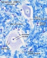
 Spinal cord histology 01.jpg 480 × 600; 116 KB
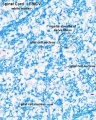
 Spinal cord histology 02.jpg 480 × 600; 121 KB
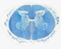
 Spinal cord histology 03.jpg 480 × 600; 103 KB
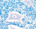
 Spinal cord histology 04.jpg 480 × 600; 119 KB
 Spinal cord histology 05.jpg 1,280 × 1,024; 463 KB
 Spinal cord histology 06.jpg 1,280 × 1,024; 318 KB
 Spinal cord histology 07.jpg 1,280 × 1,024; 365 KB
 Spinal cord histology 08.jpg 1,280 × 1,024; 418 KB
 Spinal cord histology 09.jpg 1,280 × 1,024; 227 KB


