Category:Human
From Embryology
This Embryology category shows pages and media related to human development.
Subcategories
This category has the following 72 subcategories, out of 72 total.
C
- Carnegie Embryo
- Carnegie Embryo 1
- Carnegie Embryo 112
- Carnegie Embryo 1134B
- Carnegie Embryo 116
- Carnegie Embryo 1266
- Carnegie Embryo 1455
- Carnegie Embryo 148
- Carnegie Embryo 172
- Carnegie Embryo 19
- Carnegie Embryo 239
- Carnegie Embryo 2393
- Carnegie Embryo 240
- Carnegie Embryo 248
- Carnegie Embryo 256
- Carnegie Embryo 296
- Carnegie Embryo 3527
- Carnegie Embryo 3956
- Carnegie Embryo 4046
- Carnegie Embryo 4059
- Carnegie Embryo 407
- Carnegie Embryo 4148
- Carnegie Embryo 43
- Carnegie Embryo 4405
- Carnegie Embryo 460
- Carnegie Embryo 463
- Carnegie Embryo 523
- Carnegie Embryo 5541
- Carnegie Embryo 5609
- Carnegie Embryo 5652
- Carnegie Embryo 5682
- Carnegie Embryo 5874
- Carnegie Embryo 6032
- Carnegie Embryo 625
- Carnegie Embryo 6426
- Carnegie Embryo 6581
- Carnegie Embryo 7618
- Carnegie Embryo 7669
- Carnegie Embryo 786
- Carnegie Embryo 808
- Carnegie Embryo 8147
- Carnegie Embryo 8239
- Carnegie Embryo 8370
- Carnegie Embryo 858
- Carnegie Embryo 8630
- Carnegie Embryo 8967
- Carnegie Embryo 9296
- Carnegie Embryo 96
- Carnegie Embryo 963
- Carnegie Embryo 966
- Carnegie Embryo 9697
D
F
H
Pages in category 'Human'
The following 124 pages are in this category, out of 332 total.
(previous page) (next page)P
- Paper - Models of the pancreas in embryos of the pig, rabbit, cat, and man (1908)
- Paper - Notes on the development of the human sphenoid (1910)
- Paper - Notes on the postnatal growth of the heart kidneys liver and spleen in man (1919)
- Paper - Observations on the neural crest of a ten-somite human embryo (1939)
- Paper - On The Age Of Human Embryos
- Paper - On the Development of the Human Heart
- Paper - On the Development, Ossification, and Growth of the Palate Bone of Man
- Paper - On the Frequency of Localized Anomalies in Human Embryos and Infants at Birth
- Paper - Phases of Maturation and Fertilization in Human Ova
- Paper - Significant superficial anastomoses in the arterial blood supply to the human brain (1959)
- Paper - Some Gross Structural and Quantitative Aspects of the Developmental Anatomy of the Human Embryonic, Fetal and Circumnatal Skeleton
- Paper - Some Observations on the Development of the Ventral Pancreas in Man
- Paper - Teratogenecity in the setting of cardiac development and maldevelopment
- Paper - Teratological studies (1919)
- Paper - The Anatomy of Human Embryos with Seventeen to Twenty-three Pairs of Somites
- Paper - The cartilaginous skull of a human embryo twenty-one millimeters in length (1920)
- Paper - The cortex of the brain in the human embryo during the fourth month with special reference to the so-called Papilla of Retzius
- Paper - The critical period in the development of the intestines (1914)
- Paper - The development and reduction of the tail and of the caudal end of the spinal cord (1920)
- Paper - The Development of Head-Process and Prochordal Plate in Man
- Paper - The development of synovial joints
- Paper - The Development of the Cranial and Spinal Nerves in the Occipital Region of the Human Embryo
- Paper - The development of the human prostate gland with reference to the development of other structures at the neck of the urinary bladder (1912)
- Paper - The development of the mucous membrane oesophagus stomach and small intestine in human embryo
- Paper - The development of the nervous tissues of the human embryo (1877)
- Paper - The Development of the Nose and of the Pharynx and its Derivatives in Man
- Paper - The Development of the Scala Tympani, Scala Vestibuli and Perioticular Cistern in the Human Embryo
- Paper - The development of the subcutaneous vascular plexus in the head of the human embryo (1923)
- Paper - The developmental alterations in the vascular system of the brain of the human embryo (1921)
- Paper - The Earliest Blood-Vessels in Man
- Paper - The Early Development of Man, with Special Reference to the Development of the Mesoderm and Cloacal Membrane
- Paper - The early development of the otic vesicle in staged human embryos
- Paper - The early relation of the auditory vesicle to the ectoderm in human embryos
- Paper - The Factors Involved in the Excavation of the Cavities in the Cartilaginous Capsule of the Ear in the Human Embryo
- Paper - The first appearance of the neural tube and optic primordium in the human embryo at stage 10
- Paper - The formation of the umbilical cord and the umbilical region of the anterior abdominal wall
- Paper - The genesis and structure of the membrana tectoria and the crista spiralis of the cochlea (1918)
- Paper - The Internal Genital Organs of a Female Foetus of 15 cm Length
- Paper - The Maturation of the Human Ovum
- Paper - The Origin of the Otic and Optic Primordia in Man
- Paper - The Peripheral Nervous System in the Human Embryo at the End of the First Month (10 mm)
- Paper - The prenatal development of the human temporomandibular joint
- Paper - The relations of the somites of the head to the brain in a human embryo of 20 paired somites
- Paper - The sexual differences of the fetal pelvis
- Paper - The sexual differences of the fetal pelvis (1899)
- Paper - The subdivisions of the neural folds in man
- Paper - The vascular drainage of the endolymphatic sac and its topographical relation to the transverse sinus in the human
- Paper - Transformation of the aortic-arch system during the development of the human embryo (1922)
- Paper - Two Early Human Embryos
- Paper - Two presomite human embryos
- Paper - Vertebral Regional Determination in Young Human Embryos
- Paper The development of the subcutaneous vascular plexus in the head of the human embryo (1923)
- Patent Ductus Venosus Movie
- Template:Placental villi timeline
- Template:PMID22437671 links
- Pregnancy Test
R
- Template:Ref-BascomOsterud1925
- Template:Ref-Baumgartner1917
- Template:Ref-Berry1900
- Template:Ref-Bolk1915
- Template:Ref-Carey1919
- Template:Ref-ClarkGJ1900
- Template:Ref-Gladstone1935
- Template:Ref-HertigAdams1967
- Template:Ref-Hines1921
- Template:Ref-Jirasek1980
- Template:Ref-Johnston1914
- Template:Ref-Noback1943 figures
- Template:Ref-OtisBrent1954
- Template:Ref-Romanes1941b
- Template:Ref-Schmidt1877
- Template:Ref-Schultz1919
- Template:Ref-Siddiqi1934
- Template:Ref-Valentin1834
- Template:Ref-Veit1918
S
- Template:Second trimester timeline
- SH Lecture - Lymphatic Structure and Organs
- Template:Shoulder timeline
- Template:Shoulder Timeline table2
- Template:SlideCircumvallatePlacenta
- Template:SlideOvaryCorpusLuteum
- Template:SlidePlacentalVilli1
- Template:Smell timeline
- Template:Spaulding1922
- Template:Species Placenta collapsetable1
- Template:Species Placenta table1
- Template:Spleen timeline
- Template:Spleen Timeline Table
- Template talk:Spleen Timeline Table
- Template:Stage 11 BF images
- Template:Stage19 bf2 links
- Template:Stage23oralcavity images
- Template:Streeter1908 figures
- Template:Streeter1917
- Template:Streeter1917 figures
- Template:Streeter1917a
- Template:Streeter1919 figures
- Template:Streeter1921
- Template:Sudler1902 figures
T
U
V
W
Media in category 'Human'
The following 200 files are in this category, out of 2,421 total.
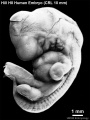
(previous page) (next page)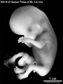 HillH13 Fetus bf01.jpg 1,200 × 1,600; 171 KB
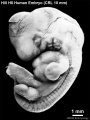
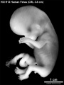 HillH13 Fetus bf02.jpg 1,200 × 1,600; 170 KB
 HillH13 Fetus.gif 450 × 600; 274 KB
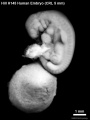
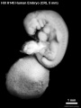 HillH145 Stage 13 bf01.jpg 908 × 1,210; 81 KB
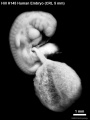
 HillH145 Stage 13 bf02.jpg 908 × 1,210; 83 KB
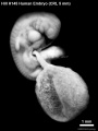
 HillH145 Stage 13 bf03.jpg 908 × 1,210; 86 KB
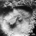
 HillH145 Stage 13 bf04.jpg 908 × 1,210; 84 KB
 HillH145 Stage 13 bf05.jpg 1,257 × 1,257; 240 KB
 HillH145 Stage 13 bf06.jpg 1,257 × 1,257; 244 KB
 HillH145 Stage 13 bf07.jpg 1,257 × 1,257; 244 KB
 HillH145 Stage 13 bf08.jpg 1,257 × 1,257; 224 KB
 HillH159 Stage 8 bf01.jpg 1,807 × 1,768; 237 KB
 HillH159 Stage 8 bf02.jpg 1,807 × 1,768; 264 KB
 HillH159 Stage 8 bf03.jpg 1,706 × 1,643; 168 KB
 HillH159 Stage 8 bf04.jpg 1,706 × 1,643; 214 KB
 HillH202 Stage 17 500.gif 500 × 500; 251 KB
 HillH202 Stage 17 600.gif 600 × 600; 346 KB
 HillH202 Stage 17 bf01.jpg 1,200 × 1,166; 173 KB
 HillH202 Stage 17 bf02.jpg 800 × 800; 85 KB
 HillH202 Stage 17 bf03.jpg 800 × 800; 86 KB
 HillH202 Stage 17 bf04.jpg 1,000 × 1,000; 138 KB
 HillH4 Stage 12 bf01.jpg 841 × 1,121; 75 KB
 HillH4 Stage 12 bf02.jpg 841 × 1,121; 75 KB
 HillH5 Stage 16 bf01.jpg 1,125 × 1,500; 142 KB
 HillH5 Stage 16 bf02.jpg 1,125 × 1,500; 142 KB
 HillH5 Stage 16 bf03.jpg 1,125 × 1,500; 148 KB
 HillH5 Stage 16 bf04.jpg 1,125 × 1,500; 149 KB
 HillH5 Stage 16 bf05.jpg 1,125 × 1,500; 116 KB
 HillH5 Stage 16 bf06.jpg 1,125 × 1,500; 111 KB
 HillH5 Stage 16 bf07.jpg 1,125 × 1,500; 151 KB
 HillH5 Stage 16 bf08.jpg 1,125 × 1,500; 149 KB
 HillH5 Stage 16 bf09.jpg 1,125 × 1,500; 109 KB
 HillH5 Stage 16 bf10.jpg 1,125 × 1,500; 110 KB
 HillH5 Stage 16 bf11.jpg 1,125 × 1,500; 147 KB
 HillH5 Stage 16 bf12.jpg 1,125 × 1,500; 154 KB
 HillH5 Stage 16 bf13.gif 450 × 600; 296 KB
 HillH5 Stage 16 bf14.gif 450 × 600; 300 KB
 HillH5 Stage 16 bf15.gif 450 × 600; 184 KB
 HillH5 Stage 16 bf16.gif 450 × 600; 257 KB
 HillH5 Stage 16 bf17.gif 450 × 600; 238 KB
 HillH5 Stage 16 bf18.gif 450 × 600; 314 KB
 HillH52 chorionic villi 01.jpg 1,200 × 924; 182 KB
 HillH52 chorionic villi 02.jpg 1,200 × 900; 155 KB
 HillH52 chorionic villi 03.jpg 1,200 × 891; 280 KB
 HillH52 chorionic villi 04.jpg 1,200 × 900; 254 KB
 HillH52 chorionic villi 05.jpg 1,200 × 992; 371 KB
 HillH52 chorionic villi 06.jpg 1,200 × 900; 346 KB
 HillH52 chorionic villi 07.jpg 1,200 × 900; 544 KB
 HillH52 chorionic villi 08.jpg 1,200 × 900; 229 KB
 HillH52 chorionic villi 09.jpg 1,200 × 900; 237 KB
 HillH52 chorionic villi 10.jpg 1,200 × 900; 253 KB
 HillH58 Stage 17 bf01.jpg 1,500 × 1,500; 273 KB
 HillH58 Stage 17 bf02.jpg 1,500 × 1,500; 279 KB
 HillH6 Stage 18 bf01.jpg 1,547 × 2,062; 274 KB
 HillH6 Stage 18 bf02.jpg 1,547 × 2,062; 337 KB
 HillH6 Stage 18 bf03.jpg 1,547 × 2,062; 287 KB
 HillH6 Stage 18 bf04.jpg 1,547 × 2,062; 318 KB
 HillH6 Stage 18 bf05.jpg 1,547 × 2,062; 218 KB
 HillH6 Stage 18 bf06.jpg 1,547 × 2,062; 232 KB
 HillH6 Stage 18 bf51.jpg 1,547 × 2,062; 198 KB
 HillH8 Stage 16 bf01.jpg 1,125 × 1,500; 136 KB
 HillH8 Stage 16 bf02.jpg 1,125 × 1,500; 145 KB
 HillH8 Stage 16 bf03.jpg 1,125 × 1,500; 170 KB
 HillH8 Stage 16 bf04.jpg 1,125 × 1,500; 171 KB
 HillH8 Stage 16 bf05.jpg 1,125 × 1,500; 176 KB
 HillH8 Stage 16 bf06.jpg 1,125 × 1,500; 169 KB
 HillH8 Stage 16 bf07.gif 600 × 800; 490 KB
 HillH8 Stage 16 bf08.gif 450 × 600; 295 KB
 HillH8 Stage 16 bf09.gif 600 × 800; 571 KB
 HillH8 Stage 16 bf10.gif 450 × 600; 338 KB
 HillH8 Stage 16 bf11.gif 600 × 800; 585 KB
 HillH8 Stage 16 bf12.gif 450 × 600; 348 KB
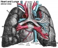 Historic-lungs.jpg 600 × 493; 79 KB
 Hochstadter 1919.jpg 845 × 1,200; 0 bytes
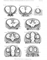
 Hochstadter plate 01.jpg 1,408 × 2,000; 255 KB
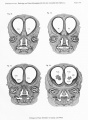
 Hochstadter plate 02.jpg 1,408 × 2,000; 305 KB
 Hochstadter plate 03.jpg 2,000 × 1,408; 258 KB
 Hochstadter plate 04.jpg 1,408 × 2,000; 273 KB
 Hochstadter plate 05.jpg 1,408 × 2,000; 235 KB
 Hochstadter plate 06.jpg 1,408 × 2,000; 280 KB
 Hochstadter plate 07.jpg 1,626 × 2,000; 261 KB
 Hochstadter plate 08.jpg 1,670 × 2,000; 367 KB
 Hochstadter plate 09.jpg 1,692 × 2,000; 307 KB
 Hochstadter plate 10.jpg 1,626 × 2,000; 551 KB
 Hochstadter plate 11.jpg 1,603 × 2,000; 570 KB
 Hochstadter plate 12.jpg 1,541 × 2,000; 543 KB
 Hochstadter plate 13.jpg 1,461 × 2,000; 531 KB
 Hochstadter plate 14.jpg 1,517 × 2,000; 574 KB
 Hochstadter plate 15.jpg 1,462 × 2,000; 450 KB
 Hochstadter plate 16.jpg 1,507 × 2,000; 640 KB
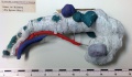 Hubrecht model-Homo16 4mm.jpg 1,707 × 1,000; 311 KB
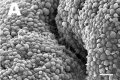 Human - uterine epithelium SEM01.jpg 600 × 400; 37 KB
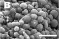 Human - uterine epithelium SEM02.jpg 600 × 400; 26 KB
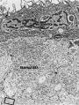
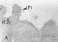 Human - uterine epithelium TEM01.jpg 600 × 433; 25 KB
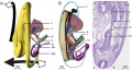
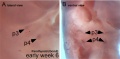
 Human 15 weeks - terminal nerve and vomeronasal organ nerves.jpg 940 × 403; 306 KB
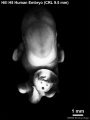
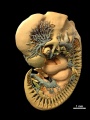
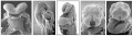
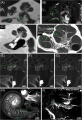
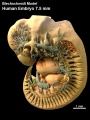 Human 7.5mm embryo model 01.jpg 747 × 1,000; 149 KB
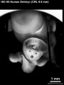
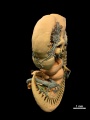
 Human 7.5mm embryo model 02.jpg 747 × 1,000; 130 KB
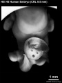
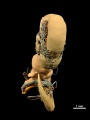
 Human 7.5mm embryo model 03.jpg 747 × 1,000; 72 KB
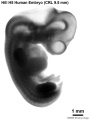
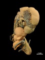
 Human 7.5mm embryo model 04.jpg 747 × 1,000; 56 KB
 Human 7.5mm embryo model 05.jpg 747 × 1,000; 87 KB
 Human 7.5mm embryo model 06.jpg 747 × 1,000; 139 KB
 Human 7.5mm embryo model 07.jpg 747 × 1,000; 118 KB
 Human 7.5mm embryo model 08.jpg 747 × 1,000; 69 KB
 Human 7.5mm embryo model 09.jpg 747 × 1,000; 63 KB
 Human 7.5mm embryo model 10.jpg 747 × 1,000; 106 KB
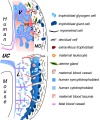 Human and mouse fetal-maternal interface cartoon.jpg 600 × 720; 91 KB
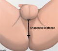 Human anogenital distance.jpg 570 × 499; 23 KB
 Human blastocyst derived stem cells.jpg 1,200 × 825; 198 KB
 Human blastocyst formation-in vitro.jpg 753 × 157; 23 KB
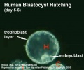 Human blastocyst hatching movie icon.jpg 498 × 414; 29 KB
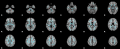 Human brain white matter tracts.png 1,200 × 490; 289 KB
 Human cochlea fetal development cartoon.jpg 592 × 1,200; 96 KB
 Human congenital diaphragmatic hernia.jpg 800 × 626; 86 KB
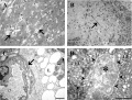 Human corpus luteum - light-and-electron-micrograph.jpg 936 × 711; 208 KB
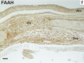
 Human developing lung protein 01.jpg 657 × 1,000; 241 KB
 Human developing lung protein 02.jpg 800 × 302; 82 KB
 Human development 001.mov ; 2 MB
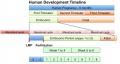
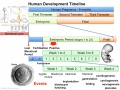 Human development timeline graph 01.jpg 1,000 × 750; 141 KB
 Human development timeline graph 02.jpg 800 × 424; 61 KB
 Human development timeline graph icon.jpg 250 × 188; 16 KB
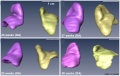
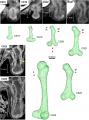 Human embryo femur CS18 to CS23.png 1,200 × 1,624; 1.42 MB
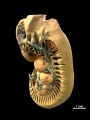
 Human embryo midgut loop 01.jpg 1,423 × 771; 200 KB
 Human embryo neck 01.jpg 534 × 827; 186 KB
 Human embryo neck 02.jpg 529 × 825; 167 KB
 Human embryo neck 03.jpg 558 × 795; 184 KB
 Human embryo parathyroid 01.jpg 1,000 × 495; 63 KB
 Human embryo skin 24 week EGA.jpg 596 × 939; 165 KB
 Human embryo skin 8-9 week EGA desmosomes.jpg 800 × 198; 40 KB
 Human embryo skin 8-9 week EGA.jpg 657 × 872; 188 KB
 Human embryo skin 9-11 week EGA.jpg 623 × 804; 176 KB
 Human Embryo Stage18-19.jpg 600 × 392; 17 KB
 Human embryo thymus and parathyroid 01.jpg 639 × 1,000; 58 KB
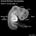 Human embryo tomography Carnegie stage 17.jpg 516 × 516; 35 KB
 Human embryonic shoulder girdle 01.jpg 1,000 × 726; 81 KB
 Human embryonic shoulder girdle 02.jpg 1,025 × 713; 109 KB
 Human embryonic shoulder girdle 04.jpg 1,000 × 755; 71 KB
 Human embryonic stem cell defined conditions 01.jpg 1,200 × 1,132; 372 KB
 Human embryonic stem cell defined conditions 02.jpg 1,091 × 411; 187 KB
 Human embryonic stem cell defined conditions 03.jpg 500 × 376; 73 KB
 Human embryonic tongue 01.jpg 1,200 × 818; 443 KB
 Human embryonic tongue 02.jpg 650 × 850; 220 KB
 Human embryonic tongue 03.jpg 650 × 470; 159 KB
 Human embryonic tongue 04.jpg 650 × 470; 131 KB
 Human embryonic tongue 05.jpg 650 × 470; 146 KB
 Human embryonic tongue 06.jpg 650 × 470; 99 KB
 Human embryonic tongue 07.jpg 650 × 470; 187 KB
 Human embryonic tongue 08.jpg 650 × 470; 185 KB
 Human embryonic tongue 09.jpg 650 × 470; 180 KB
 Human embryonic tongue 10.jpg 650 × 470; 118 KB
 Human embryonic tongue 11.jpg 650 × 470; 155 KB
 Human embryonic-fetal tongue 01.jpg 1,000 × 1,129; 490 KB
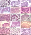
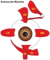 Human extraocular muscles 01.jpg 500 × 600; 47 KB
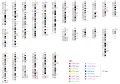
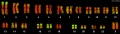 Human female karyotype 01.jpg 2,000 × 562; 118 KB
 Human fertilization movie 1 frame 01.jpg 600 × 409; 27 KB
 Human fertilization movie 1 frame 02.jpg 600 × 409; 27 KB
 Human fertilization movie 1 frame 03.jpg 600 × 409; 26 KB
 Human fertilization movie 1 frame 04.jpg 600 × 409; 24 KB
 Human fertilization movie 1 frame 05.jpg 600 × 409; 25 KB
 Human fertilization movie 1 frame 06.jpg 600 × 409; 25 KB
 Human fertilization movie 1 frame 07.jpg 600 × 409; 24 KB
 Human fertilization movie 1 frame 08.jpg 600 × 409; 25 KB
 Human fertilization movie 1 frame 09.jpg 600 × 409; 24 KB
 Human fertilization movie 1 frame 10.jpg 600 × 409; 25 KB
 Human fetal adrenal GA32 large.mp4 ; 2.62 MB
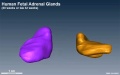 Human fetal adrenal GA32.jpg 800 × 502; 39 KB
 Human fetal adrenal gland 01.jpg 1,266 × 800; 107 KB
 Human fetal cochlea 01.jpg 1,270 × 532; 266 KB
 Human fetal cochlea 02.jpg 1,270 × 532; 271 KB
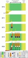
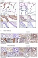
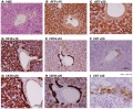
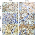 Human fetal gonad retinoid receptor expression.jpg 1,004 × 1,000; 447 KB
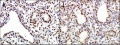
 Human fetal kidney histology 01.jpg 1,280 × 1,024; 481 KB
 Human fetal kidney histology 02.jpg 1,280 × 1,024; 322 KB
 Human fetal kidney histology 03.jpg 1,280 × 1,024; 333 KB
 Human fetal kidney histology 04.jpg 1,280 × 1,024; 307 KB
 Human fetal membrane 01.jpg 726 × 545; 69 KB
 Human fetal membrane 02.jpg 726 × 545; 87 KB
 Human fetal membrane 03.jpg 726 × 545; 88 KB
 Human fetal neck 01.jpg 549 × 827; 186 KB
 Human fetal neural aneuploidy.jpg 1,000 × 1,400; 134 KB
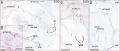
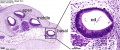
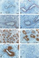
 Human fetal ovary SMAD6 expression.jpg 711 × 535; 167 KB
 Human fetal tongue 01.jpg 1,500 × 672; 350 KB
 Human fetal uterus myometrium.jpg 500 × 554; 86 KB
 Human heart developmental functional networks.jpg 833 × 614; 424 KB
 Human heart SEM1.jpg 1,200 × 330; 47 KB
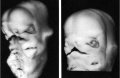
 Human holoprosencephaly cyclopia dissection.jpg 600 × 340; 37 KB
 Human homeobox genes.jpg 1,200 × 840; 222 KB
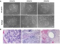 Human induced pluripotent stem cells 01.jpg 1,091 × 799; 250 KB
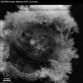
 Human inner ear MicroCT.jpg 2,131 × 3,111; 1,001 KB
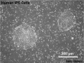 Human iPS cells 01.jpg 598 × 448; 83 KB
 Human liver week 9.jpg 1,200 × 991; 425 KB
 Human lung pseudoglandular.jpg 672 × 1,000; 121 KB
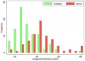 Human male anogenital distance graph.jpg 600 × 429; 28 KB
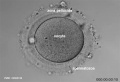
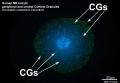 Human MII oocyte 01.jpg 1,200 × 826; 92 KB
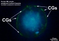 Human MII oocyte 02.jpg 1,200 × 826; 98 KB
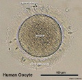
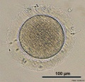 Human oocyte 01.jpg 700 × 675; 98 KB
 Human oocyte 11.jpg 700 × 675; 104 KB
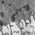 Human oocyte em01.jpg 600 × 589; 65 KB
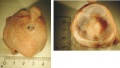
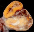
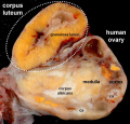
 Human ovary - corpus luteum 01.jpg 1,024 × 979; 162 KB
 Human ovary - corpus luteum 02.jpg 837 × 800; 119 KB
 Human ovary - corpus luteum 11.jpg 1,024 × 979; 89 KB


