File:Mouse E18 neurovasculature MicroCT.jpg
Original file (960 × 364 pixels, file size: 68 KB, MIME type: image/jpeg)
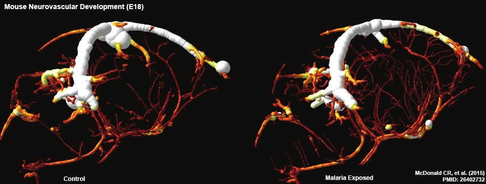
Mouse E18 Cerebral Vasculature MicroCT
Complement activation, in particular C5a, may contribute to neuropathology and adverse outcomes during malaria in pregnancy. Animal model results show that experimental malaria in pregnancy nduces neurocognitive deficits in offspring and suggest novel targets for intervention.
- Links: Mouse E18 Cerebral Vasculature MicroCT | Abnormal Development - Malaria | Computed Tomography | Developmental Origins of Health and Disease | Mouse Development
Reference
<pubmed>26402732</pubmed>| PLoS Pathog.
Copyright
© 2015 McDonald et al. This is an open access article distributed under the terms of the Creative Commons Attribution License, which permits unrestricted use, distribution, and reproduction in any medium, provided the original author and source are credited
Original image adjusted in size and labelling.
Cite this page: Hill, M.A. (2026, October 4) Embryology Mouse E18 neurovasculature MicroCT.jpg. Retrieved from https://embryology.med.unsw.edu.au/embryology/index.php/File:Mouse_E18_neurovasculature_MicroCT.jpg
- © Dr Mark Hill 2026, UNSW Embryology ISBN: 978 0 7334 2609 4 - UNSW CRICOS Provider Code No. 00098G
File history
Yi efo/eka'e gwa ebo wo le nyangagi wuncin ye kamina wunga tinya nan
| Gwalagizhi | Nyangagi | Dimensions | User | Comment | |
|---|---|---|---|---|---|
| current | 12:51, 28 September 2015 | | 960 × 364 (68 KB) | Z8600021 (talk | contribs) | ==Mouse E18 Cerebral Vasculature MicroCT== Complement activation, in particular C5a, may contribute to neuropathology and adverse outcomes during malaria in pregnancy. Animal model results show that experimental malaria in pregnancy nduces neurocogniti... |
You cannot overwrite this file.
File usage
The following 2 pages use this file:


