File:Mesoderm-cartoon4.jpg
From Embryology
No higher resolution available.
Mesoderm-cartoon4.jpg (400 × 300 pixels, file size: 20 KB, MIME type: image/jpeg)
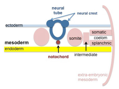
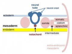
Mesoderm and Ectoderm Development cartoon
This final simplified cartoon in the cross-section view development series shows:
- Mesoderm - lateral plate mesoderm division into somatic and splanchnic mesoderm separated by intra-embryonic coelom.
- Ectoderm - neural plate has now formed the neural tube and neural crest separation from the overlying epithelial ectoderm.
Simplified Cartoon of Mesoderm and Ectoderm Development (week 3-4)
- Mesoderm and Ectoderm Cartoons
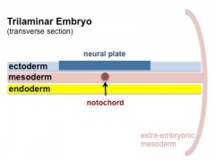
Trilaminar Embryo
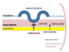
Paraxial and Lateral Plate
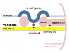
Somites
Somatic and Splanchnic


- Mesoderm Links: 1 Trilaminar Embryo | 2 Paraxial and Lateral Plate | 3 Somites | 4 Somatic and Splanchnic | Animation | Mesoderm | Somitogenesis | Notochord
Cite this page: Hill, M.A. (2026, October 6) Embryology Mesoderm-cartoon4.jpg. Retrieved from https://embryology.med.unsw.edu.au/embryology/index.php/File:Mesoderm-cartoon4.jpg
- © Dr Mark Hill 2026, UNSW Embryology ISBN: 978 0 7334 2609 4 - UNSW CRICOS Provider Code No. 00098G
File history
Yi efo/eka'e gwa ebo wo le nyangagi wuncin ye kamina wunga tinya nan
| Gwalagizhi | Nyangagi | Dimensions | User | Comment | |
|---|---|---|---|---|---|
| current | 17:04, 16 Mayıs 2014 | | 400 × 300 (20 KB) | Z8600021 (talk | contribs) |
You cannot overwrite this file.
File usage
The following 51 pages use this file:
- 2017ANAT2341 Tutorial 2
- BGDA Lecture - Development of the Embryo/Fetus 1
- BGDA Lecture - Development of the Embryo/Fetus 2
- BGDA Lecture - Development of the Nervous System
- BGDA Practical 3 - Notochord
- BGDA Practical 3 - Week 3 Summary
- BGDA Practical 7 - Week 3
- BGDA Practical 7 - Week 4
- BGDB Face and Ear - Trilaminar Embryo
- BGDB Gastrointestinal - Activity 1
- BGDB Gastrointestinal - Trilaminar Embryo
- BGD Lecture - Gastrointestinal System Development
- BGD Lecture - Sexual Differentiation
- Coelomic Cavity - Abnormalities
- Coelomic Cavity Development
- Gastrointestinal Tract - Oesophagus Development
- I
- Lecture - Early Vascular Development
- Lecture - Ectoderm Development
- Lecture - Gastrointestinal Development
- Lecture - Genital Development
- Lecture - Limb Development
- Lecture - Mesoderm Development
- Lecture - Musculoskeletal Development
- Mesoderm
- REI - Reproductive Medicine Seminar 2018
- Renal System Development
- Royal Hospital for Women - Reproductive Medicine Seminar 2018
- Somitogenesis
- Splanchnic Mesoderm
- File:Mesoderm-cartoon1.jpg
- File:Mesoderm-cartoon2.jpg
- File:Mesoderm-cartoon3.jpg
- File:Mesoderm-cartoon4.jpg
- File:Mesoderm cartoon.gif
- File:Mesoderm cartoon1.gif
- File:Mesoderm cartoon2.gif
- File:Mesoderm cartoon3.gif
- File:Mesoderm cartoon4.gif
- File:Mesoderm cartoon 01.jpg
- File:Mesoderm cartoon 02.jpg
- File:Mesoderm cartoon 03.jpg
- File:Mesoderm cartoon 04.jpg
- File:Stage10 K12202-01.jpg
- File:Stage10 K12202-02.jpg
- File:Stage10 K12202-03.jpg
- File:Stage10 K12202-04.jpg
- File:Stage10 K12202-05.jpg
- File:Stage10 K12202-06.jpg
- Template:Mesoderm cartoon
- Template:Stage10 K12202 images


