File:Mouse meiosis pachytene 01.jpg
From Embryology
Size of this preview: 800 × 198 pixels. Other resolution: 1,280 × 317 pixels.
Original file (1,280 × 317 pixels, file size: 50 KB, MIME type: image/jpeg)
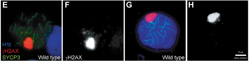
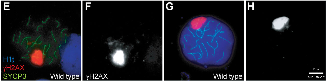
Spermatocytes activate a recombination-dependent arrest at pachynema
- blue - H1t (mid/late pachytene stage marker)
- red - γH2AX (sex body)
- green - SYCP3 (synaptonemal complex component)
| Cell Division Links: meiosis | mitosis | Lecture - Cell Division and Fertilization | spermatozoa | oocyte | fertilization | zygote | Genetics |
Reference
PMID 25768017
Copyright
© 2015 Pacheco et al. This is an open access article distributed under the terms of the Creative Commons Attribution License, which permits unrestricted use, distribution, and reproduction in any medium, provided the original author and source are credited
Fig 1. http://dx.doi.org/10.1371/journal.pgen.1005017.g001
Cite this page: Hill, M.A. (2026, October 8) Embryology Mouse meiosis pachytene 01.jpg. Retrieved from https://embryology.med.unsw.edu.au/embryology/index.php/File:Mouse_meiosis_pachytene_01.jpg
- © Dr Mark Hill 2026, UNSW Embryology ISBN: 978 0 7334 2609 4 - UNSW CRICOS Provider Code No. 00098G
File history
Yi efo/eka'e gwa ebo wo le nyangagi wuncin ye kamina wunga tinya nan
| Gwalagizhi | Nyangagi | Dimensions | User | Comment | |
|---|---|---|---|---|---|
| current | 20:03, 15 Mayıs 2016 | | 1,280 × 317 (50 KB) | Z8600021 (talk | contribs) | ==Spermatocytes activate a recombination-dependent arrest at pachynema== (A) Apoptotic Trip13mod/mod spermatocyte showing the axial element protein SYCP3 (green), TUNEL (green), mid/late pachytene stage marker H1t (blue) and γH2AX staining (red). Abs... |
You cannot overwrite this file.
File usage
The following page uses this file: