File:Zebrafish day 1 SEM.jpg
Original file (1,000 × 750 pixels, file size: 130 KB, MIME type: image/jpeg)
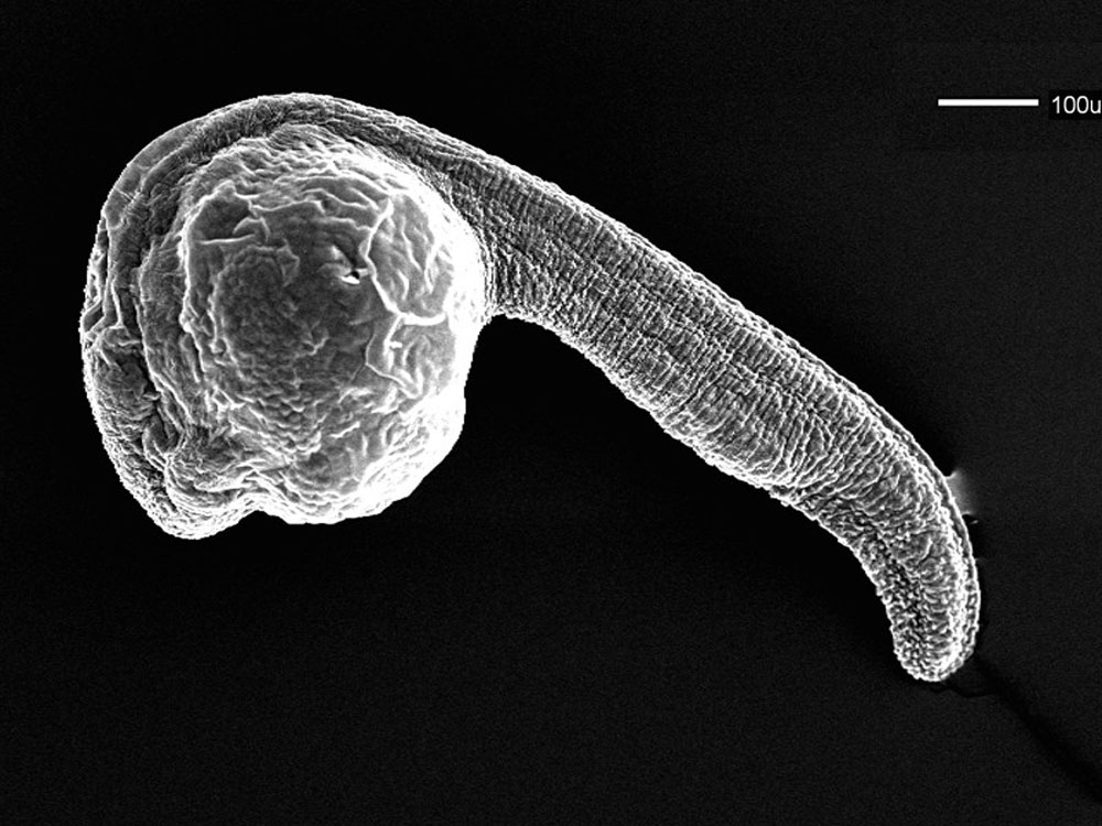
Zebrafish Day 1 SEM
Scanning EM of a 24 hr (prim-5) zebrafish embryo.
- Links: Image - day 1 | Image - brain fold | Image - myotomes | Image - trunk | Image - trunk | Image - perichordal sheath | Image - enveloping layer | Image - enveloping layer | Zebrafish Development | Scanning Electron Microscopy
Image Source: Scanning electron micrographs of the Zebrafish embryos are reproduced with the permission of Associate Professor Bryan Crawford, Department of Biology, University of New Brunswick.
Reference
Zebrafish were chemically fixed critically point dried, and sputter coated with gold/palladium. This image is part of a series taken by Bryan Crawford while he was at the University of Washington. They are part of the Zebrafish--The Living Laboratory CD made available by Mark Cooper and described in Methods in Cell Biology Volume 77, 2004, Pages 439-457.
Original image name: 12661.jpg http://www.cellimagelibrary.org/images/12661
Copyright
Licensing: Attribution Non-Commercial Share Alike:This image is licensed under a Creative Commons Attribution, Non-Commercial Share Alike License.
File history
Yi efo/eka'e gwa ebo wo le nyangagi wuncin ye kamina wunga tinya nan
| Gwalagizhi | Nyangagi | Dimensions | User | Comment | |
|---|---|---|---|---|---|
| current | 08:16, 26 April 2011 | | 1,000 × 750 (130 KB) | S8600021 (talk | contribs) | ==Zebrafish Day 1 SEM== Scanning EM of a 24 hr (prim-5) zebrafish embryo. Zebrafish were chemically fixed critically point dried, and sputter coated with gold/palladium. This image is part of a series taken by Bryan Crawford while he was at the Univer |
You cannot overwrite this file.
File usage
The following page uses this file: