File:Keibel Mall 2 166.jpg
Original file (969 × 1,000 pixels, file size: 144 KB, MIME type: image/jpeg)
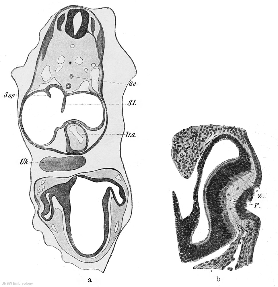
Fig. 166 a and b. Human embryo of 5 mm
a: Section through the anterior part of the head and through the heart region of a human embryo of 5 mm. X 25.
b: Section through the anlage of an eye of the same embryo. X 100.
(After drawings by Hammar, from the Normentafel of Keibel and Elze.)
F., thread-like tissue between the lens and the anlage of the retina; Oe., oesophagus; SI, septum I (Born); S.sp., septum spurium (His); Tr.a., truncus arteriosus; Uk., lower jaw; Z., cell mass in the lens pit.
| Embryology - 24 Jun 2024 |
|---|
| Google Translate - select your language from the list shown below (this will open a new external page) |
|
العربية | català | 中文 | 中國傳統的 | français | Deutsche | עִברִית | हिंदी | bahasa Indonesia | italiano | 日本語 | 한국어 | မြန်မာ | Pilipino | Polskie | português | ਪੰਜਾਬੀ ਦੇ | Română | русский | Español | Swahili | Svensk | ไทย | Türkçe | اردو | ייִדיש | Tiếng Việt These external translations are automated and may not be accurate. (More? About Translations) |
Felix W. The development of the urinogenital organs. In Keibel F. and Mall FP. Manual of Human Embryology II. (1912) J. B. Lippincott Company, Philadelphia. pp 752-979.
| Historic Disclaimer - information about historic embryology pages |
|---|
 |
Cite this page: Hill, M.A. (2024, June 24) Embryology Keibel Mall 2 166.jpg. Retrieved from https://embryology.med.unsw.edu.au/embryology/index.php/File:Keibel_Mall_2_166.jpg
- © Dr Mark Hill 2024, UNSW Embryology ISBN: 978 0 7334 2609 4 - UNSW CRICOS Provider Code No. 00098G
File history
Yi efo/eka'e gwa ebo wo le nyangagi wuncin ye kamina wunga tinya nan
| Gwalagizhi | Nyangagi | Dimensions | User | Comment | |
|---|---|---|---|---|---|
| current | 09:53, 21 February 2014 | | 969 × 1,000 (144 KB) | Z8600021 (talk | contribs) | ==Fig. 166 a and b. Human embryo of 5 mm== a: Section through the anterior part of the head and through the heart region of a human embryo of 5 mm. X 25. b: Section through the anlage of an eye of the same embryo. X 100. (After drawings by Hammar, ... |
You cannot overwrite this file.
File usage
The following page uses this file: