File:Fetal head medial.jpg
Fetal_head_medial.jpg (632 × 447 pixels, file size: 34 KB, MIME type: image/jpeg)
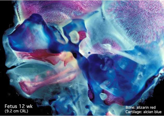
Fetal Head (12 weeks)
Medial view, stained for bone (Histology_Stains#Alizarin_Redalizarin red) and cartilage (alcian blue)
In this medial (internal) view, note the distribution of cartilage from the nasal region through the base of the skull to the atlas/axis (with new bone forming). See also the original Meckel's cartilage within the newly forming bony mandible.
Note: Cartilage in the neck where the hyoid bone will eventually form. Meckel's cartilage visible in the medial view within the newly forming bone.
- Links: Fetal Development - 12 Weeks | Week 12 - Head histology | Week 12 - Head medial cartilage/bone | Week 12 - Head lateral cartilage/bone | Head Development | Gastrointestinal Tract Development | Bone Development
Original File Name: Headbone2.jpg
Source: Prof Virginia Diewert
Cite this page: Hill, M.A. (2026, October 10) Embryology Fetal head medial.jpg. Retrieved from https://embryology.med.unsw.edu.au/embryology/index.php/File:Fetal_head_medial.jpg
- © Dr Mark Hill 2026, UNSW Embryology ISBN: 978 0 7334 2609 4 - UNSW CRICOS Provider Code No. 00098G
File history
Yi efo/eka'e gwa ebo wo le nyangagi wuncin ye kamina wunga tinya nan
| Gwalagizhi | Nyangagi | Dimensions | User | Comment | |
|---|---|---|---|---|---|
| current | 16:45, 13 September 2009 | | 632 × 447 (34 KB) | S8600021 (talk | contribs) |
You cannot overwrite this file.
File usage
The following 40 pages use this file:
- 2009 Lecture 13
- 2010 BGD Lecture - Development of the Embryo/Fetus 2
- 2010 BGD Practical 12 - Second Trimester
- 2010 BGD Practical 6 - Week 7
- 2010 Foundations Lecture - Introduction to Human Development
- 2010 Lab 6
- 2010 Lecture 13
- 2011 Lab 12 - Second Trimester
- 2011 Lab 6 - Fetal
- AACP Meeting 2013 - Face Embryology
- ANAT2241 Bone, Bone Formation and Joints
- ANAT2341 Lab 11 - Embryo to Fetus
- ANAT2341 Lab 12 - Second Trimester
- ANAT2341 Lab 6 - Fetal
- Abnormal Development - Fetal Growth Restriction
- BGDA Lecture - Development of the Embryo/Fetus 2
- BGDA Practical 12 - Second Trimester
- BGDA Practical 7 - Week 7
- BGDB Face and Ear - Fetal
- BGD Lecture - Face and Ear Development
- Bone Development
- Bone Histology
- Cartilage Histology
- Fetal Development
- Fetal Development - 12 Weeks
- Foundations Lecture - Introduction to Human Development
- Head Development
- Joint Development - Temporomandibular Joint
- Lecture - Fetal Development
- Lecture - Head Development
- Lecture - Musculoskeletal Development
- Musculoskeletal System - Abnormalities
- Musculoskeletal System - Axial Skeleton Development
- Musculoskeletal System - Bone Development
- Musculoskeletal System - Cartilage Development
- Musculoskeletal System - Joint Development
- Musculoskeletal System - Skull Development
- Musculoskeletal System Development
- Pre-Medicine Program - Embryology
- Virginia Diewert


