File:Brain ventricles and ganglia development 03.jpg
Original file (989 × 583 pixels, file size: 36 KB, MIME type: image/jpeg)
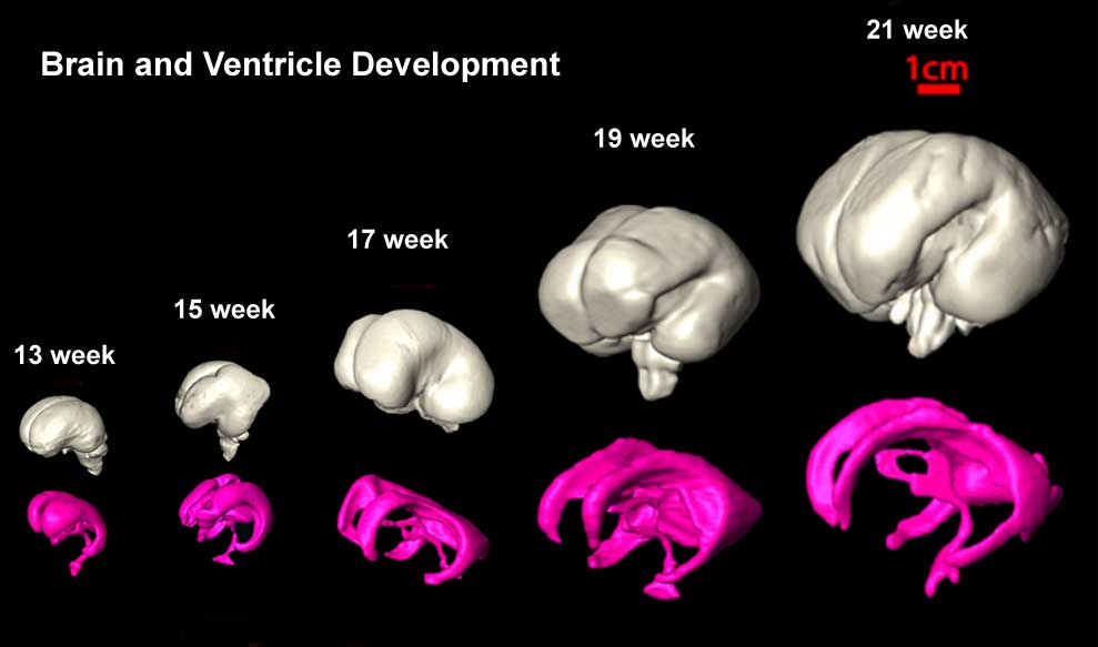
Brain and Ventricle Development
Three-dimensional reconstruction of the basal ganglia and ganglionic eminence (bottom row of a), ventricle (middle row of a), and whole brain (top row of a).
Different colors represent different brain structures: whole brain (gray), ventricle (pink), ganglionic eminence (red), putamen and globus pallidus together (cyan), thalamus (yellow), and caudate nucleus (green).
Original File Name: Figure 9 Original image modified by scaling relative to the Week 21 brain.
Diffusion tensor imaging (DTI) A newly developed form of magnetic resonance imaging (MRI). Magnetic field variations of the MRI magnet are applied in at least six different directions generating a three dimensional shape of the diffusion pattern. This technique can be used in neural imaging of white matter due to the orientation of axon bundles and the associated water flow. (More? Magnetic Resonance Imaging)
- Neural DTI Links: Scaled Fissures 13-21 weeks | Fissures 13-21 weeks | Brain Sylvian Fissure | Scaled Brain and Ventricles 13-21 weeks | Scaled Brain, Ventricles and Ganglia 13-21 weeks | Limbic Tract 13-19 weeks | Brain and Ventricles 13-21 weeks | Sylvian Fissure Movie | Neural System Development | Magnetic Resonance Imaging
Reference
<pubmed>19339620</pubmed>| PMC2721010 | J Neurosci.
Copyright: Copyright of all material published in The Journal of Neuroscience remains with the authors. The authors grant the Society for Neuroscience an exclusive license to publish their work for the first 6 months. After 6 months the work becomes available to the public to copy, distribute, or display under a Creative Commons Attribution-Noncommercial-Share Alike 3.0 Unported license.
File history
Yi efo/eka'e gwa ebo wo le nyangagi wuncin ye kamina wunga tinya nan
| Gwalagizhi | Nyangagi | Dimensions | User | Comment | |
|---|---|---|---|---|---|
| current | 14:46, 27 August 2010 | | 989 × 583 (36 KB) | S8600021 (talk | contribs) | ==Brain and Ventricle Development== Three-dimensional reconstruction of the basal ganglia and ganglionic eminence (bottom row of a), ventricle (middle row of a), and whole brain (top row of a). Different colors represent different brain structures: whol |
You cannot overwrite this file.
File usage
The following 17 pages use this file:
- 2010 Lecture 23
- 2014 Group Project 7
- ANAT2341 Lab 10 - Fetal
- BGDA Lecture - Development of the Nervous System
- BGDA Practical 12 - Second Trimester
- Brain Awareness Week 2012
- K12 Brain Awareness Week
- Lecture - Fetal Development
- Lecture - Neural Development
- M
- Magnetic Resonance Imaging
- Neural - Cerebrum Development
- Neural - Ventricular System Development
- Neural System - Fetal
- Neural System - Postnatal
- Neural System Development
- Talk:2014 Group Project 7